

Human P16 Primary Antibody
Item Information
Catalog #
Size
Price
Description
p16 (cyclin-dependent kinase inhibitor 2A, INK4a) is a tumor suppressor protein. It is a specific inhibitor of Cdk 4 / Cdk 6, and a tumor suppressor involved in the pathogenesis of a variety of malignancies. Recent analyses of the p16 INK4a gene revealed homozygous deletions, nonsense, missense, or frameshift mutations in several human cancers. Although the frequency of p16 INK4a abnormalities is higher in tumor derived cell lines than in unselected primary tumors, significant subsets of clinical cases with aberrant p16 INK4a gene have been reported among melanomas, gliomas, esophageal, pancreatic, lung, and urinary bladder carcinomas, and some types of leukemia.
Product Overview
Entrez GenelD
1029
Aliases
Human P16
Clone#
5A8A4; 3G8D12
Host / Isotype
Mouse / IgG1
Species Reactivity
Human
Immunogen
Purified recombinant fragment of P16 expressed in E. Coli.
Formulation
Ascitic fluid containing 0.03% sodium azide.
Storage
Store at 4°C short term. Aliquot and store at -20°C long term. Avoid freeze/thaw cycles.
Product Applications
WB (Western Blot)
1/500 - 1/2000
IHC_P(Immunohistochemistry)
1/200 - 1/1000
ELISA
1/10000
References
1. Bai, F. et al. Mol. Cell. Biol.2003 23, 1269-1277.
2. Lowe, S.W. and Sherr, C.J. Curr. Opin. Genet.2003 Dev.13, 77-83.
3. Sherr, C.J. Nat. Rev. Mol. Cell Biol.2001 2, 731-737.
2. Lowe, S.W. and Sherr, C.J. Curr. Opin. Genet.2003 Dev.13, 77-83.
3. Sherr, C.J. Nat. Rev. Mol. Cell Biol.2001 2, 731-737.
Product Image
Western Blot
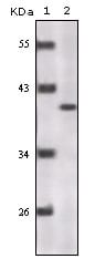
Figure 1: Western blot analysis using P16 mouse mAb against truncated P16 recombinant protein.
Immunohistochemical analysis

Figure 2: Immunohistochemical analysis of paraffin-embedded human lung adenocarcinoma (A), esophageal squamous cell carcinoma (B), hepatic cell carcinoma (C), thyroid tumor (D), breast adenofibroma (E), breast infiltrating ductal carcinoma (F), normal cerebrum tissue (G), normal colon tissue (H), normal esophageal tissue (I), showing nuclear localization using P16 mouse mAb with DAB staining.
Immunohistochemical analysis

Figure 3: Immunohistochemical analysis of paraffin-embedded human spleen tissues using P16 mouse mAb.
For Research Use Only. Not for use in diagnostic procedures.