

HTR3A Primary Antibody
Item Information
Catalog #
Size
Price
Description
The product of this gene belongs to the ligand-gated ion channel receptor superfamily. This gene encodes subunit A of the type 3 receptor for 5-hydroxytryptamine (serotonin), a biogenic hormone that functions as a neurotransmitter, a hormone, and a mitogen. This receptor causes fast, depolarizing responses in neurons after activation. It appears that the heteromeric combination of A and B subunits is necessary to provide the full functional features of this receptor, since either subunit alone results in receptors with very low conductance and response amplitude. Alternatively spliced transcript variants encoding different isoforms have been identified.
Product Overview
Entrez GenelD
3359
Aliases
HTR3; 5HT3R; 5-HT-3; 5-HT3A; 5-HT3R
Clone#
4D5E7
Host / Isotype
Mouse / IgG2a
Species Reactivity
Human
Immunogen
Purified recombinant fragment of human HTR3A (AA: extra 24-157) expressed in E. Coli.
Formulation
Purified antibody in PBS with 0.05% sodium azide
Storage
Store at 4°C short term. Aliquot and store at -20°C long term. Avoid freeze/thaw cycles.
Product Applications
WB (Western Blot)
1/500 - 1/2000
IHC_P(Immunohistochemistry)
1/200 - 1/1000
ICC (Immunocytochemistry)
1/100 - 1/500
FCM (Flow Cytometry)
1/200 - 1/400
ELISA
1/10000
References
1.PLoS One. 2015 Dec 23;10(12):e0145269.
2.Iran J Allergy Asthma Immunol. 2014 Feb;13(1):33-9.
2.Iran J Allergy Asthma Immunol. 2014 Feb;13(1):33-9.
Product Image
Elisa

Figure 1: Black line: Control Antigen (100 ng);Purple line: Antigen (10ng); Blue line: Antigen (50 ng); Red line:Antigen (100 ng)
Western Blot

Figure 2:Western blot analysis using HTR3A mAb against human HTR3A (AA: extra 24-157) recombinant protein. (Expected MW is 41.9 kDa)
Western Blot

Figure 3:Western blot analysis using HTR3A mAb against HEK293 (1) and HTR3A (AA: extra 24-157)-hIgGFc transfected HEK293 (2) cell lysate.
Immunofluorescence analysis
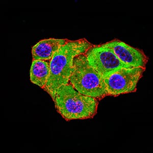
Figure 4:Immunofluorescence analysis of Hela cells using HTR3A mouse mAb (green). Blue: DRAQ5 fluorescent DNA dye. Red: Actin filaments have been labeled with Alexa Fluor- 555 phalloidin. Secondary antibody from Fisher (Cat#: 35503)
Flow cytometric

Figure 5:Flow cytometric analysis of Hela cells using HTR3A mouse mAb (green) and negative control (red).
Immunohistochemical analysis
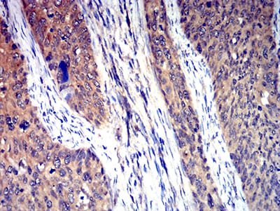
Figure 6:Immunohistochemical analysis of paraffin-embedded cervical cancer tissues using HTR3A mouse mAb with DAB staining.
For Research Use Only. Not for use in diagnostic procedures.