

HSPA9 Primary Antibody
Item Information
Catalog #
Size
Price
Description
This gene encodes a member of the heat shock protein 70 gene family. The encoded protein is primarily localized to the mitochondria but is also found in the endoplasmic reticulum, plasma membrane and cytoplasmic vesicles. This protein is a heat-shock cognate protein. This protein plays a role in cell proliferation, stress response and maintenance of the mitochondria. A pseudogene of this gene is found on chromosome 2.
Product Overview
Entrez GenelD
3313
Aliases
CSA; MOT; MOT2; SAAN; CRP40; EVPLS; GRP75; PBP74; GRP-75; HSPA9B; SIDBA4; MTHSP75; HEL-S-124m
Clone#
3A12G3
Host / Isotype
Mouse / Mouse IgG1
Species Reactivity
Human, Monkey, Rat
Immunogen
Purified recombinant fragment of human HSPA9 (AA: 480-679) expressed in mammalian.
Formulation
Purified antibody in PBS with 0.05% sodium azide
Storage
Store at 4°C short term. Aliquot and store at -20°C long term. Avoid freeze/thaw cycles.
Product Applications
WB (Western Blot)
1/500 - 1/2000
IHC_P(Immunohistochemistry)
1/200 - 1/1000
ICC (Immunocytochemistry)
1/200 - 1/1000
FCM (Flow Cytometry)
1/200 - 1/400
ELISA
1/10000
References
1,Cancer Lett . 2021 Apr 1;502:25-33.
2,Exp Mol Pathol . 2021 Feb;118:104593.
2,Exp Mol Pathol . 2021 Feb;118:104593.
Product Image
Elisa
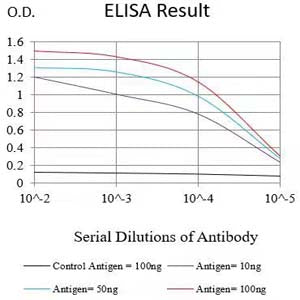
Figure 1:Black line: Control Antigen (100 ng);Purple line: Antigen (10ng); Blue line: Antigen (50 ng); Red line:Antigen (100 ng)
Western Blot
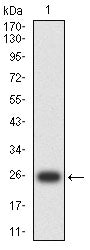
Figure 2:Western blot analysis using HSPA9 mAb against human HSPA9 (AA: 480-679) recombinant protein. (Expected MW is 25.2 kDa)
Western Blot

Figure 3:Western blot analysis using HSPA9 mouse mAb against CSO-7 (1),C6 (2), PC-12 (3), PANC-1(4),A549 (5),MCF-7 (6),K562 (7),Hela (8),A431 (9),HepG2 (10)and Jurkat (11) cell lysate.
Immunohistochemical analysis

Figure 4:Immunofluorescence analysis of Hela cells using HSPA9 mouse mAb (green). Blue: DRAQ5 fluorescent DNA dye. Red: Actin filaments have been labeled with Alexa Fluor- 555 phalloidin. Secondary antibody from Fisher (Cat#: 35503)
Immunofluorescence analysis

Figure 5:Flow cytometric analysis of Hela cells using HSPA9 mouse mAb (green) and negative control (red).
Immunofluorescence analysis

Figure 6:Flow cytometric analysis of HepG2 cells using HSPA9 mouse mAb (green) and negative control (red).
Immunofluorescence analysis

Figure 7:Flow cytometric analysis of Jurkat cells using HSPA9 mouse mAb (green) and negative control (red).
Immunohistochemical analysis

Figure 8:Immunohistochemical analysis of paraffin-embedded cervical carcinoma tissues using HSPA9 mouse mAb with DAB staining.
Immunohistochemical analysis

Figure 9:Immunohistochemical analysis of paraffin-embedded ovarian cancer tissues using HSPA9 mouse mAb with DAB staining.
For Research Use Only. Not for use in diagnostic procedures.