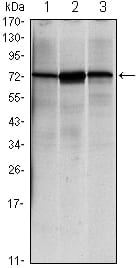
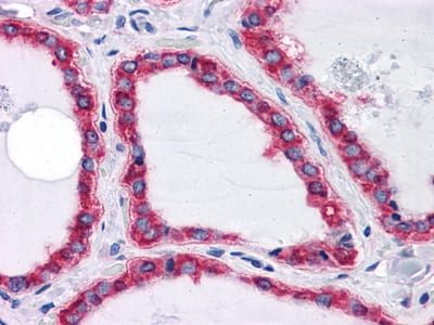
HSPA5 Primary Antibody
When Chinese hamster K12 cells are starved of glucose, the synthesis of several proteins, called glucose-regulated proteins (GRPs), is markedly increased. Hendershot et al. (1994) (PubMed 8020977) pointed out that one of these, GRP78 (HSPA5), also referred to as 'immunoglobulin heavy chain-binding protein' (BiP), is a member of the heat-shock protein-70 (HSP70) family and is involved in the folding and assembly of proteins in the endoplasmic reticulum (ER). Because so many ER proteins interact transiently with GRP78, it may play a key role in monitoring protein transport through the cell.Probably plays a role in facilitating the assembly of multimeric protein complexes inside the ER.The HSP70 proteins are ubiquitous molecular chaparones that are found in all organisms and tissue types. Like other members of the HSP70 family, BiP is a peptide-binding ATPase that is able to differentiate native proteins from unfolded polypeptides. BiP does not bind to fully folded and assembled proteins, except in the presence of other co-chaparones. BiP is involved in a number of key mechanisms and pathways including polypeptide translocation across the endoplasmic reticulum, folding, assembly, transport of secreted or membrane proteins, and the regulation of calcium homeostasis. Although BiP is relatively abundant, marked increases in BiP occur where there is an accumulation of unfolded polypeptides. For this reason, BiP has been identified as a marker for various disease states that are associated with secretory and transmembrane protein misfolding.
2. J Virol. 2009 Dec;83(23):12622-5.
3. Mod Pathol. 2010 Jan;23(1):45-53.