

Mouse Monoclonal Antibody to HSP70
Item Information
Catalog #
Size
Price
Description
HSPA4 (Heat Shock Protein Family A (Hsp70) Member 4) is a Protein Coding gene. Diseases associated with HSPA4 include Vulvovaginitis and Babesiosis. Among its related pathways are Cellular response to heat stress and Mechanisms of CFTR activation by S-nitrosoglutathione (normal and CF). An important paralog of this gene is HSPA4L.
Product Overview
Entrez GenelD
3308
Aliases
RY; APG-2; HSPH2; hsp70; hsp70RY; HEL-S-5a; HS24/P52
Clone#
2E4F10B11
Host / Isotype
Mouse / IgG1
Immunogen
Purified recombinant fragment of human HSP70 (AA: 642-841) expressed in mammalian.
Formulation
Purified antibody in PBS with 0.05% sodium azide
Storage
Store at 4°C short term. Aliquot and store at -20°C long term. Avoid freeze/thaw cycles.
Product Applications
WB (Western Blot)
1/500 - 1/2000
IHC_P(Immunohistochemistry)
1/200 - 1/1000
ICC (Immunocytochemistry)
1/200 - 1/1000
FCM (Flow Cytometry)
1/200 - 1/400
ELISA
1/10000
References
1.J Biol Chem.2020 Jun 12;295(24):8302-8324.2.Biol Chem.2020 Oct 25;401(11):1233-1248.
Product Image
Elisa

Figure 1:Black line: Control Antigen (100 ng);Purple line: Antigen (10ng); Blue line: Antigen (50 ng); Red line:Antigen (100 ng)
Western Blot

Figure 3:Western blot analysis using HSP70 mouse mAb against Hela (1), HepG2 (2),Hek293 (3),COS-7 (4),A549 (5) and Jurkat (6) cell lysate.
Immunofluorescence analysis

Figure 4:Immunofluorescence analysis of Hela cells using HSP70 mouse mAb (green). Blue: DRAQ5 fluorescent DNA dye. Red: Actin filaments have been labeled with Alexa Fluor- 555 phalloidin. Secondary antibody from Fisher (Cat#: 35503)
Flow cytometric analysis

Figure 5:Flow cytometric analysis of Jurkat cells using HSP70 mouse mAb (green) and negative control (red).
Flow cytometric analysis

Figure 6:Flow cytometric analysis of K562 cells using HSP70 mouse mAb (green) and negative control (red).
Flow cytometric analysis

Figure 7:Flow cytometric analysis of Raji cells using HSP70 mouse mAb (green) and negative control (red).
Immunohistochemical analysis
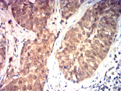
Figure 8:Immunohistochemical analysis of paraffin-embedded bladder cancer tissues using HSP70 mouse mAb with DAB staining.
Western Blot

Figure 9:Western blot analysis using HSP70 mAb against human HSP70 (AA: 642-841) recombinant protein. (Expected MW is 53.3 kDa)
Immunohistochemical analysis

Figure 9:Immunohistochemical analysis of paraffin-embedded rectal cancer tissues using HSP70 mouse mAb with DAB staining.
For Research Use Only. Not for use in diagnostic procedures.