

HOXA9 Primary Antibody
Item Information
Catalog #
Size
Price
Description
In vertebrates, the genes encoding the class of transcription factors called homeobox genes are found in clusters named A, B, C, and D on four separate chromosomes. Expression of these proteins is spatially and temporally regulated during embryonic development. This gene is part of the A cluster on chromosome 7 and encodes a DNA-binding transcription factor which may regulate gene expression, morphogenesis, and differentiation. This gene is highly similar to the abdominal-B (Abd-B) gene of Drosophila. A specific translocation event which causes a fusion between this gene and the NUP98 gene has been associated with myeloid leukemogenesis. Read-through transcription exists between this gene and the upstream homeobox A10 (HOXA10) gene.
Product Overview
Entrez GenelD
3205
Aliases
HOX1; ABD-B; HOX1G; HOX1.7
Clone#
5C7C6
Host / Isotype
Mouse / IgG2a
Species Reactivity
Human
Immunogen
Purified recombinant fragment of human HOXA9 (AA: 1-272) expressed in E. Coli.
Formulation
Purified antibody in PBS with 0.05% sodium azide
Storage
Store at 4°C short term. Aliquot and store at -20°C long term. Avoid freeze/thaw cycles.
Product Applications
WB (Western Blot)
1/500 - 1/2000
ICC (Immunocytochemistry)
1/200 - 1/1000
FCM (Flow Cytometry)
1/200 - 1/400
ELISA
1/10000
References
1.BMC Cancer. 2014 May 21;14:353.
2.Oncol Res. 2013;20(10):467-72.
2.Oncol Res. 2013;20(10):467-72.
Product Image
Elisa

Figure 1: Black line: Control Antigen (100 ng); Purple line: Antigen(10ng); Blue line: Antigen (50 ng); Red line: Antigen (100 ng);
Western Blot

Figure 2:Western blot analysis using HOXA9 mAb against human HOXA9 (AA: 1-272) recombinant protein. (Expected MW is 56.1 kDa)
Western Blot

Figure 3:Western blot analysis using HOXA9 mAb against HEK293 (1) and HOXA9 (AA: 1-272)-hIgGFc transfected HEK293 (2) cell lysate.
Immunofluorescence analysis
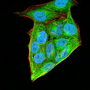
Figure 4:Immunofluorescence analysis of Hela cells using HOXA9 mouse mAb (green). Blue: DRAQ5 fluorescent DNA dye. Red: Actin filaments have been labeled with Alexa Fluor- 555 phalloidin. Secondary antibody from Fisher (Cat#: 35503)
Flow cytometric

Figure 5:Flow cytometric analysis of Hela cells using HOXA9 mouse mAb (green) and negative control (red).
For Research Use Only. Not for use in diagnostic procedures.