

Mouse Monoclonal Antibody to HMOX1
Item Information
Catalog #
Size
Price
Description
Heme oxygenase, an essential enzyme in heme catabolism, cleaves heme to form biliverdin, which is subsequently converted to bilirubin by biliverdin reductase, and carbon monoxide, a putative neurotransmitter. Heme oxygenase activity is induced by its substrate heme and by various nonheme substances. Heme oxygenase occurs as 2 isozymes, an inducible heme oxygenase-1 and a constitutive heme oxygenase-2. HMOX1 and HMOX2 belong to the heme oxygenase family.
Product Overview
Entrez GenelD
3162
Aliases
HO-1; HSP32; HMOX1D; bK286B10
Clone#
2B2A1
Host / Isotype
Mouse / IgG1
Immunogen
Purified recombinant fragment of human HMOX1 (AA: 1-110) expressed in E. Coli.
Formulation
Purified antibody in PBS with 0.05% sodium azide
Storage
Store at 4°C short term. Aliquot and store at -20°C long term. Avoid freeze/thaw cycles.
Product Applications
WB (Western Blot)
1/500 - 1/2000
IHC_P(Immunohistochemistry)
1/200 - 1/1000
FCM (Flow Cytometry)
1/200 - 1/400
ELISA
1/10000
References
1.Sci Rep. 2020 Oct 28;10(1):18506.2.Cells. 2020 Oct 15;9(10):2298.
Product Image
Elisa
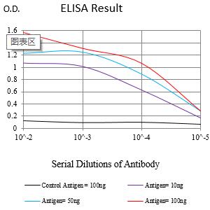
Figure 1:Black line: Control Antigen (100 ng);Purple line: Antigen (10ng); Blue line: Antigen (50 ng); Red line:Antigen (100 ng)
Western Blot

Figure 2:Western blot analysis using HMOX1 mAb against human HMOX1 (AA:1-110 ) recombinant protein. (Expected MW is 38.8 kDa)
Western Blot

Figure 3:Western blot analysis using HMOX1 mAb against HEK293-6e (1) and HMOX1 (AA: 1-110)-hIgGFc transfected HEK293-6e (2) cell lysate.
Flow cytometric analysis
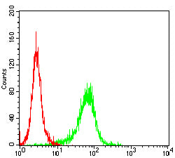
Figure 4:Flow cytometric analysis of Jurkat cells using HMOX1 mouse mAb (green) and negative control (red).
Immunohistochemical analysis

Figure 5:Immunohistochemical analysis of paraffin-embedded Liver tissues using HMOX1 mouse mAb with DAB staining.
Immunohistochemical analysis

Figure 6:Immunohistochemical analysis of paraffin-embedded liver cancer tissues using HMOX1 mouse mAb with DAB staining.
For Research Use Only. Not for use in diagnostic procedures.