HLA-DRB1

Item Information
Catalog #
Size
Price
Description

HLA-DRB1 belongs to the HLA class II beta chain paralogs. The class II molecule is a heterodimer consisting of an alpha (DRA) and a beta chain (DRB), both anchored in the membrane. It plays a central role in the immune system by presenting peptides derived from extracellular proteins. Class II molecules are expressed in antigen presenting cells. The beta chain is approximately 26-28 kDa. It is encoded by 6 exons. Exon one encodes the leader peptide; exons 2 and 3 encode the two extracellular domains; exon 4 encodes the transmembrane domain; and exon 5 encodes the cytoplasmic tail. Within the DR molecule the beta chain contains all the polymorphisms specifying the peptide binding specificities. Hundreds of DRB1 alleles have been described and some alleles have increased frequencies associated with certain diseases or conditions. For example, DRB1*1302 has been related to acute and chronic hepatitis B virus persistence. There are multiple pseudogenes of this gene.

Product Overview
Entrez GenelD
3123
Aliases
SS1; DRB1; HLA-DRB; HLA-DR1B
Clone#
5B3D2
Host / Isotype
Mouse / Mouse IgG1
Species Reactivity
Human
Immunogen
Purified recombinant fragment of human HLA-DRB1 (AA: 30-266) expressed in E. Coli.
Formulation
Purified antibody in PBS with 0.05% sodium azide
Storage
4℃; -20℃ for long term storage
Product Applications
WB (Western Blot)
1/500 - 1/2000
IHC_P(Immunohistochemistry)
1/200 - 1/1000
FCM (Flow Cytometry)
1/200 - 1/400
ELISA
1/10000
References
1.Front Immunol. 2021 Jul 15;12:691475.
2.Ann Lab Med. 2022 Jan 1;42(1):54-62.
Product Image
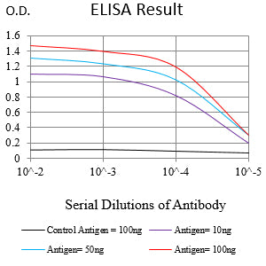
Elisa
Figure 1:Black line: Control Antigen (100 ng);Purple line: Antigen (10ng); Blue line: Antigen (50 ng); Red line:Antigen (100 ng)
Western Blot
Figure 2:Western blot analysis using HLA-DRB1 mAb against human HLA-DRB1 (AA: 30-266) recombinant protein. (Expected MW is 53.7 kDa)
Western Blot
Figure 3:Western blot analysis using HLA-DRB1 mAb against HEK293-6e (1) and HLA-DRB1 (AA: 30-266)-hIgGFc transfected HEK293-6e (2) cell lysate.
Immunofluorescence analysis
Figure 5:Flow cytometric analysis of Raji cells using HLA-DRB1 mouse mAb (green) and negative control (red).
Immunohistochemical analysis
Figure 5:Immunohistochemical analysis of paraffin-embedded human stomach cancer tissue using HLA-DRB1 mouse mAb with DAB staining.
Immunohistochemical analysis
Figure 6:Immunohistochemical analysis of paraffin-embedded human tonsil tissue using HLA-DRB1 mouse mAb with DAB staining.
For Research Use Only. Not for use in diagnostic procedures.