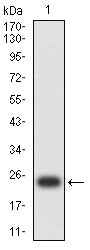
HLA-DPA1

Item Information
Catalog #
Size
Price
Description

HLA-DPA1 belongs to the HLA class II alpha chain paralogues. This class II molecule is a heterodimer consisting of an alpha (DPA) and a beta (DPB) chain, both anchored in the membrane. It plays a central role in the immune system by presenting peptides derived from extracellular proteins. Class II molecules are expressed in antigen presenting cells (APC: B lymphocytes, dendritic cells, macrophages). The alpha chain is approximately 33-35 kDa and its gene contains 5 exons. Exon one encodes the leader peptide, exons 2 and 3 encode the two extracellular domains, exon 4 encodes the transmembrane domain and the cytoplasmic tail. Within the DP molecule both the alpha chain and the beta chain contain the polymorphisms specifying the peptide binding specificities, resulting in up to 4 different molecules.

Product Overview
Entrez GenelD
3113
Aliases
DPA1; PLT1; HLADP; HLASB; DP(W3); DP(W4); HLA-DPA; HLA-DP1A; HLA-DPB1
Clone#
2B1A10
Host / Isotype
Mouse / Mouse IgG2a
Species Reactivity
Human
Immunogen
Purified recombinant fragment of human HLA-DPA1 (AA: 29-209) expressed in E. Coli.
Formulation
Purified antibody in PBS with 0.05% sodium azide
Storage
4℃; -20℃ for long term storage
Product Applications
WB (Western Blot)
1/500 - 1/2000
IHC_P(Immunohistochemistry)
1/200 - 1/1000
FCM (Flow Cytometry)
1/200 - 1/400
ELISA
1/10000
References
1.HLA. 2021 Dec;98(6):577-578.
2.HLA. 2022 Jan;99(1):74-75.
Product Image
Elisa
Figure 1:Black line: Control Antigen (100 ng);Purple line: Antigen (10ng); Blue line: Antigen (50 ng); Red line:Antigen (100 ng)
Western Blot
Figure 2:Western blot analysis using HLA-DPA1 mAb against human HLA-DPA1 (AA: 29-209) recombinant protein. (Expected MW is 23.7 kDa)
Western Blot
Figure 3:Western blot analysis using HLA-DPA1 mAb against HEK293-6e (1) and HLA-DPA1 (AA:29-209)-hIgGFc transfected HEK293-6e (2) cell lysate.
Western Blot
Figure 4:Western blot analysis using HLA-DPA1 mouse mAb against Raji(1) cell lysate.
Immunofluorescence analysis
Figure 5:Flow cytometric analysis of Jurkat cells using HLA-DPA1 mouse mAb (green) and negative control (red).
Immunohistochemical analysis
Figure 6:Immunohistochemical analysis of paraffin-embedded ovarian cancer tissues using HLA-DPA1 mouse mAb with DAB staining.
Immunohistochemical analysis
Figure 7:Immunohistochemical analysis of paraffin-embedded breast cancer tissues using HLA-DPA1 mouse mAb with DAB staining.
Immunohistochemical analysis
Figure 8:Immunohistochemical analysis of paraffin-embedded gastric cancer tissues using HLA-DPA1 mouse mAb with DAB staining.
Immunohistochemical analysis
Figure 9:Immunohistochemical analysis of paraffin-embedded rectal cancer tissues using HLA-DPA1 mouse mAb with DAB staining.
For Research Use Only. Not for use in diagnostic procedures.