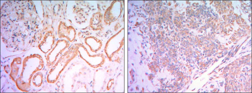
HK1 Primary Antibody

Item Information
Catalog #
Size
Price
Description

The hexokinases utilize Mg-ATP as a phosphoryl donor to catalyze the first step of intracellular glucose metabolism, the conversion of glucose to glucose- 6-phosphate. Four hexokinase isoenzymes have been identified, including hexokinase I (HXK I), hexokinase II (HXK II), hexokinase III (HXK III) and hexokinase IV (HXK IV, also designated glucokinase or GCK). Hexokinases I-III each contain an N-terminal cluster of hydrophobic amino acids. Glucokinase lacks the N-terminal hydrophobic cluster. The hydrophobic cluster is thought to be necessary for membrane binding. This is substantiated by the finding that glucokinase has lower affinity for glucose than do the other hexokinases. HK I has been shown to be expressed in brain, kidney and heart tissues as well as in hepatoma cell lines.

Product Overview
Entrez GenelD
3098
Aliases
HKI; HXK1; HK1-ta; HK1-tb; HK1-tc; HK1
Clone#
7A7
Host / Isotype
Mouse / IgG1
Species Reactivity
Human, Rat, Mouse
Immunogen
Purified recombinant fragment of human HK1 expressed in E. Coli.
Formulation
Ascitic fluid containing 0.03% sodium azide.
Storage
4°C; -20°C for long term storage
Product Applications
WB (Western Blot)
1/500 - 1/2000
IHC_P(Immunohistochemistry)
1/200 - 1/1000
ICC (Immunocytochemistry)
1/200 - 1/1000
ELISA
1/10000
References
1. Cell. 2005 Sep 23;122(6):957-68.
2. J Biomed Sci. 2007 Mar;14(2):195-202.
3. J Neural Transm. 2009 Mar;116(3):275-89.
Product Image
Western Blot
Figure 1: Western blot analysis using HK1 mouse mAb against Jurkat (1), Hela (2), HepG2 (3), MCF-7 (4) and PC-12 (5) cell lysate.
Immunohistochemical analysis
Figure 2: Immunohistochemical analysis of paraffin-embedded human salivary gland tissues (left) and kidney tissues (right) using HK1 mouse mAb with DAB staining.
Immunofluorescence analysis
Figure 3: Immunofluorescence analysis of NIH/3T3 cells using HK1 mouse mAb (green). Blue: DRAQ5 fluorescent DNA dye. Red: Actin filaments have been labeled with Alexa Fluor-555 phalloidin.
For Research Use Only. Not for use in diagnostic procedures.