

HIST2H4A(20Me) Primary Antibody
Item Information
Catalog #
Size
Price
Description
Histones are basic nuclear proteins that are responsible for the nucleosome structure of the chromosomal fiber in eukaryotes. This structure consists of approximately 146 bp of DNA wrapped around a nucleosome, an octamer composed of pairs of each of the four core histones (H2A, H2B, H3, and H4). The chromatin fiber is further compacted through the interaction of a linker histone, H1, with the DNA between the nucleosomes to form higher order chromatin structures. This gene is intronless and encodes a member of the histone H4 family. Transcripts from this gene lack polyA tails; instead, they contain a palindromic termination element. This gene is found in a histone cluster on chromosome 1. This gene is one of four histone genes in the cluster that are duplicated; this record represents the centromeric copy.
Product Overview
Entrez GenelD
8370
Aliases
H4; H4/n; H4F2; H4FN; FO108; HIST2H4
Clone#
3E7D9
Host / Isotype
Mouse / IgG1
Species Reactivity
Human
Immunogen
Synthesized peptide of human HIST2H4A ( AA: GGAKRHRK(Me)VLRDNIQ ) .
Formulation
Purified antibody in PBS with 0.05% sodium azide
Storage
Store at 4°C short term. Aliquot and store at -20°C long term. Avoid freeze/thaw cycles.
Product Applications
IHC_P(Immunohistochemistry)
1/200 - 1/1000
ICC (Immunocytochemistry)
1/200 - 1/1000
ELISA
1/10000
References
1.J Virol. 2011 Dec;85(24):13234-52.
2.Mol Cell Biol. 2003 Feb;23(4):1460-9.
2.Mol Cell Biol. 2003 Feb;23(4):1460-9.
Product Image
Elisa

Figure 1: Black line: Control Antigen (100 ng); Purple line: Antigen(10ng); Blue line: Antigen (50 ng); Red line: Antigen (100 ng);
Immunofluorescence analysis

Figure 2:Immunofluorescence analysis of HeLa cells . Blue: DRAQ5 fluorescent DNA dye. Red: Actin filaments have been labeled with Alexa Fluor- 555 phalloidin.
Immunofluorescence analysis
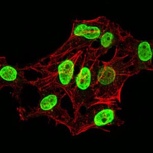
Figure 3:Immunofluorescence analysis of HeLa cells using HIST2H4A(20Me) mouse mAb (green). Blue: DRAQ5 fluorescent DNA dye. Red: Actin filaments have been labeled with Alexa Fluor- 555 phalloidin. Secondary antibody from Fisher (Cat#: 35503)
Immunohistochemical analysis

Figure 4:Immunohistochemical analysis of paraffin-embedded esophageal cancer tissues using HIST2H4A(20Me) mouse mAb with DAB staining.
Immunohistochemical analysis

Figure 5:Immunohistochemical analysis of paraffin-embedded colon cancer tissues using HIST2H4A(20Me) mouse mAb with DAB staining.
For Research Use Only. Not for use in diagnostic procedures.