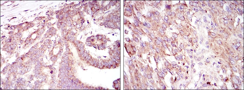
HIF1A Primary Antibody

Item Information
Catalog #
Size
Price
Description

Hypoxia-inducible factor-1 (HIF1) is a transcription factor found in mammalian cells cultured under reduced oxygen tension that plays an essential role in cellular and systemic homeostatic responses to hypoxia. HIF1 is a heterodimer composed of an alpha subunit and a beta subunit. The beta subunit has been identified as the aryl hydrocarbon receptor nuclear translocator (ARNT). This gene encodes the alpha subunit of HIF-1. Overexpression of a natural antisense transcript (aHIF) of this gene has been shown to be associated with nonpapillary renal carcinomas. Two alternative transcripts encoding different isoforms have been identified. (provided by RefSeq) Tissue specificity: Expressed in most tissues with highest levels in kidney and heart. Overexpressed in the majority of common human cancers and their metastases, due to the presence of intratumoral hypoxia and as a result of mutations in genes encoding oncoproteins and tumor suppressors.

Product Overview
Entrez GenelD
3091
Aliases
HIF1; MOP1; PASD8; bHLHe78; HIF-1alpha; HIF1-ALPHA; HIF1A
Clone#
1A3
Host / Isotype
Mouse / IgG1
Species Reactivity
Human, Mouse, Monkey
Immunogen
Purified recombinant fragment of human HIF1A expressed in E. Coli.
Formulation
Ascitic fluid containing 0.03% sodium azide.
Storage
4°C; -20°C for long term storage
Product Applications
WB (Western Blot)
1/500 - 1/2000
IHC_P(Immunohistochemistry)
1/200 - 1/1000
ICC (Immunocytochemistry)
1/200 - 1/1000
ELISA
1/10000
References
1. Int J Radiat Oncol Biol Phys. 2008 Dec 1;72(5):1551-9.
2. Eur J Appl Physiol. 2009 Mar;105(4):515-24.
Product Image
Western Blot
Figure 1: Western blot analysis using HIF1A mouse mAb against Cos7 (1), Hela (2), Jurkat (3), RAJI (4) and NIH/3T3 (5) cell lysate.
Immunohistochemical analysis
Figure 2: Immunohistochemical analysis of paraffin-embedded liver cancer tissues (left) and lung cancer tissues (right) using HIF1A mouse mAb with DAB staining.
Immunohistochemical analysis
Figure 3: Immunohistochemical analysis of paraffin-embedded stomach cancer tissues (left) and brain tumor tissues (right) using HIF1A mouse mAb with DAB staining.
Immunofluorescence analysis
Figure 4: Immunofluorescence analysis of Hela cells using HIF1A mouse mAb (green). Blue: DRAQ5 fluorescent DNA dye. Red: Actin filaments have been labeled with Alexa Fluor-555 phalloidin.
Elisa
Red: Control Antigen (100ng); Purple: Antigen (10ng); Green: Antigen (50ng); Blue: Antigen (100ng);
For Research Use Only. Not for use in diagnostic procedures.