

Mouse Monoclonal Antibody to HDAC1
Item Information
Catalog #
Size
Price
Description
Histone acetylation and deacetylation, catalyzed by multisubunit complexes, play a key role in the regulation of eukaryotic gene expression. The protein encoded by this gene belongs to the histone deacetylase/acuc/apha family and is a component of the histone deacetylase complex. It also interacts with retinoblastoma tumor-suppressor protein and this complex is a key element in the control of cell proliferation and differentiation. Together with metastasis-associated protein-2, it deacetylates p53 and modulates its effect on cell growth and apoptosis.
Product Overview
Entrez GenelD
3065
Aliases
HD1; RPD3; KDAC1; GON-10; RPD3L1
Clone#
1B6A7
Host / Isotype
Mouse / IgG1
Immunogen
Purified recombinant fragment of human HDAC1 (AA: 321-482) expressed in E. Coli.
Formulation
Purified antibody in PBS with 0.05% sodium azide
Storage
Store at 4°C short term. Aliquot and store at -20°C long term. Avoid freeze/thaw cycles.
Product Applications
WB (Western Blot)
1/500 - 1/2000
IHC_P(Immunohistochemistry)
1/200 - 1/1000
FCM (Flow Cytometry)
1/200 - 1/400
ELISA
1/10000
References
1.Cell Commun Signal. 2019 Jul 29;17(1):86. 2.Hum Pathol. 2019 Mar;85:194-201.
Product Image
Elisa
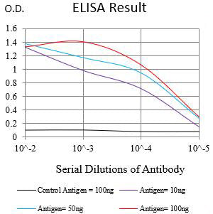
Figure 1:Black line: Control Antigen (100 ng);Purple line: Antigen (10ng); Blue line: Antigen (50 ng); Red line:Antigen (100 ng)
Western Blot
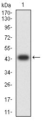
Figure 2:Western blot analysis using HDAC1 mAb against human HDAC1 (AA: 321-482) recombinant protein. (Expected MW is 44.6 kDa)
Western Blot

Figure 3:Western blot analysis using HDAC1 mAb against HEK293-6e (1) and HDAC1 (AA: 1-482)-hIgGFc transfected HEK293-6e (2) cell lysate.
Western Blot
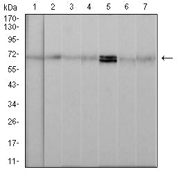
Figure 4:Western blot analysis using HDAC1 mouse mAb against NIH/3T3 (1), Hela (2), Raw264.7 (3), K562 (4), Jurkat (5), C6 (6), and Raji (7) cell lysate.
Flow cytometric analysis
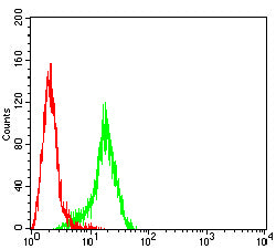
Figure 5:Flow cytometric analysis of Raji cells using HDAC1 mouse mAb (green) and negative control (red).
Immunohistochemical analysis
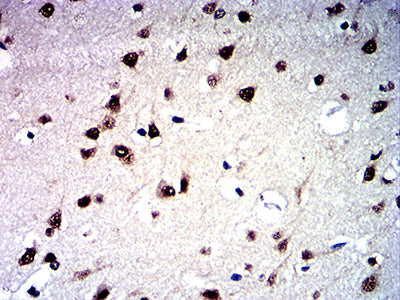
Figure 6:Immunohistochemical analysis of paraffin-embedded human brain tissues using HDAC1 mouse mAb with DAB staining.
Immunohistochemical analysis
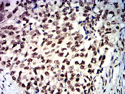
Figure 7:Immunohistochemical analysis of paraffin-embedded bladder cancer tissues using HDAC1 mouse mAb with DAB staining.
For Research Use Only. Not for use in diagnostic procedures.