

GUCY1A3 Primary Antibody
Item Information
Catalog #
Size
Price
Description
Soluble guanylate cyclases are heterodimeric proteins that catalyze the conversion of GTP to 3',5'-cyclic GMP and pyrophosphate. The protein encoded by this gene is an alpha subunit of this complex and it interacts with a beta subunit to form the guanylate cyclase enzyme, which is activated by nitric oxide. Several transcript variants encoding a few different isoforms have been found for this gene.
Product Overview
Entrez GenelD
2982
Aliases
GUCA3; GC-SA3; GUC1A3; GUCSA3; GUCY1A1
Clone#
3G6B2
Host / Isotype
Mouse / IgG1
Species Reactivity
Human
Immunogen
Purified recombinant fragment of human GUCY1A3 (AA: 22-214) expressed in E. Coli.
Formulation
Ascitic fluid containing 0.03% sodium azide.
Storage
Store at 4°C short term. Aliquot and store at -20°C long term. Avoid freeze/thaw cycles.
Product Applications
WB (Western Blot)
1/500 - 1/2000
IHC_P(Immunohistochemistry)
1/200 - 1/1000
ICC (Immunocytochemistry)
1/200 - 1/1000
FCM (Flow Cytometry)
1/200 - 1/400
ELISA
1/10000
References
1.Mol Endocrinol. 2012 Feb;26(2):292-307.
2.J Biol Inorg Chem. 2011 Dec;16(8):1227-39.
2.J Biol Inorg Chem. 2011 Dec;16(8):1227-39.
Product Image
Western Blot

Figure 1: Western blot analysis using GUCY1A3 mAb against human GUCY1A3 recombinant protein. (Expected MW is 47.2 kDa)
Western Blot

Figure 2: Western blot analysis using GUCY1A3 mAb against HEK293 (1) and GUCY1A3 (AA: 22-214)-hIgGFc transfected HEK293 (2) cell lysate.
Western Blot

Figure 3: Western blot analysis using GUCY1A3 mouse mAb against HEK293 (1) and Jurkat (2) cell lysate.
Immunofluorescence analysis

Figure 4: Immunofluorescence analysis of HepG2 cells using GUCY1A3 mouse mAb (green). Blue: DRAQ5 fluorescent DNA dye.
Flow cytometric

Figure 5: Flow cytometric analysis of HEK293 cells using GUCY1A3 mouse mAb (green) and negative control (purple).
Immunohistochemical analysis

Figure 6: Immunohistochemical analysis of paraffin-embedded renal cancer tissues using GUCY1A3 mouse mAb with DAB staining.
Immunohistochemical analysis

Figure 7: Immunohistochemical analysis of paraffin-embedded esophageal cancer tissues using GUCY1A3 mouse mAb with DAB staining.
Elisa
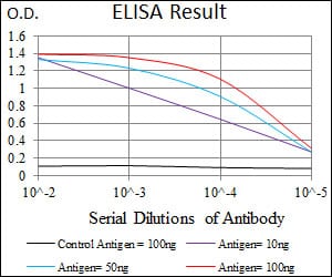
Black line: Control Antigen (100 ng); Purple line: Antigen(10ng); Blue line: Antigen (50 ng); Red line: Antigen (100 ng);
For Research Use Only. Not for use in diagnostic procedures.