

GRM3 Primary Antibody
Item Information
Catalog #
Size
Price
Description
L-glutamate is the major excitatory neurotransmitter in the central nervous system and activates both ionotropic and metabotropic glutamate receptors. Glutamatergic neurotransmission is involved in most aspects of normal brain function and can be perturbed in many neuropathologic conditions. The metabotropic glutamate receptors are a family of G protein-coupled receptors, that have been divided into 3 groups on the basis of sequence homology, putative signal transduction mechanisms, and pharmacologic properties. Group I includes GRM1 and GRM5 and these receptors have been shown to activate phospholipase C. Group II includes GRM2 and GRM3 while Group III includes GRM4, GRM6, GRM7 and GRM8. Group II and III receptors are linked to the inhibition of the cyclic AMP cascade but differ in their agonist selectivities.
Product Overview
Entrez GenelD
2913
Aliases
GLUR3; mGlu3; GPRC1C; MGLUR3
Clone#
7A5A6
Host / Isotype
Mouse / IgG2a
Species Reactivity
Human
Immunogen
Purified recombinant fragment of human GRM3 (AA: extra 433-576) expressed in E. Coli.
Formulation
Purified antibody in PBS with 0.05% sodium azide
Storage
Store at 4°C short term. Aliquot and store at -20°C long term. Avoid freeze/thaw cycles.
Product Applications
WB (Western Blot)
1/500 - 1/2000
IHC_P(Immunohistochemistry)
1/200 - 1/1000
ICC (Immunocytochemistry)
1/200 - 1/1000
FCM (Flow Cytometry)
1/200 - 1/400
ELISA
1/10000
References
1.Prog Neuropsychopharmacol Biol Psychiatry. 2015 Oct 1;62:14-21.
2.Cell Mol Biol (Noisy-le-grand). 2014 Sep 6;60(2):42-9.
2.Cell Mol Biol (Noisy-le-grand). 2014 Sep 6;60(2):42-9.
Product Image
Elisa
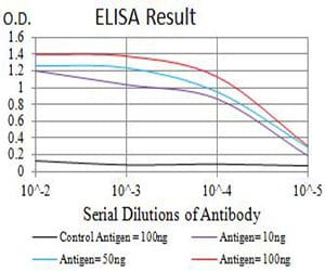
Figure 1: Black line: Control Antigen (100 ng);Purple line: Antigen (10ng); Blue line: Antigen (50 ng); Red line:Antigen (100 ng)
Western Blot
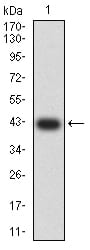
Figure 2:Western blot analysis using GRM3 mAb against human GRM3 (AA: extra 433-576) recombinant protein. (Expected MW is 42.4 kDa)
Western Blot
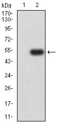
Figure 3:Western blot analysis using GRM3 mAb against HEK293 (1) and GRM3 (AA: extra 433-576)-hIgGFc transfected HEK293 (2) cell lysate.
Immunofluorescence analysis
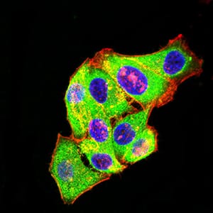
Figure 4:Immunofluorescence analysis of Hela cells using GRM3 mouse mAb (green). Blue: DRAQ5 fluorescent DNA dye. Red: Actin filaments have been labeled with Alexa Fluor- 555 phalloidin. Secondary antibody from Fisher (Cat#: 35503)
Flow cytometric
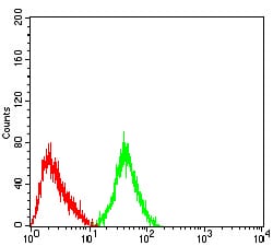
Figure 5:Flow cytometric analysis of SH-SY5Y cells using GRM3 mouse mAb (green) and negative control (red).
Immunohistochemical analysis
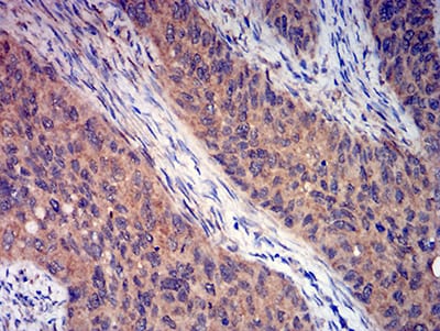
Figure 6:Immunohistochemical analysis of paraffin-embedded cervical cancer tissues using GRM3 mouse mAb with DAB staining.
For Research Use Only. Not for use in diagnostic procedures.