

GRIK4 Primary Antibody
Item Information
Catalog #
Size
Price
Description
This gene encodes a protein that belongs to the glutamate-gated ionic channel family. Glutamate functions as the major excitatory neurotransmitter in the central nervous system through activation of ligand-gated ion channels and G protein-coupled membrane receptors. The protein encoded by this gene forms functional heteromeric kainate-preferring ionic channels with the subunits encoded by related gene family members. Alternatively spliced transcript variants encoding different isoforms have been found for this gene.
Product Overview
Entrez GenelD
2900
Aliases
KA1; EAA1; GRIK; GluK4
Clone#
8H5H9
Host / Isotype
Mouse / IgG2b
Species Reactivity
Human
Immunogen
Purified recombinant fragment of human GRIK4 (AA: extra 21-166) expressed in E. Coli.
Formulation
Purified antibody in PBS with 0.05% sodium azide
Storage
Store at 4°C short term. Aliquot and store at -20°C long term. Avoid freeze/thaw cycles.
Product Applications
WB (Western Blot)
1/500 - 1/2000
IHC_P(Immunohistochemistry)
1/200 - 1/1000
ICC (Immunocytochemistry)
1/200 - 1/1000
FCM (Flow Cytometry)
1/200 - 1/400
ELISA
1/10000
References
1.Pharmacogenomics. 2014 Aug;15(11):1451-9.
2.Am J Med Genet B Neuropsychiatr Genet. 2012 Jan;159B(1):21-9.
2.Am J Med Genet B Neuropsychiatr Genet. 2012 Jan;159B(1):21-9.
Product Image
Elisa
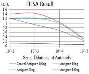
Figure 1: Black line: Control Antigen (100 ng);Purple line: Antigen (10ng); Blue line: Antigen (50 ng); Red line:Antigen (100 ng)
Western Blot
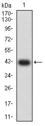
Figure 2:Western blot analysis using GRIK4 mAb against human GRIK4 (AA: extra 21-166) recombinant protein. (Expected MW is 42 kDa)
Western Blot
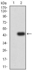
Figure 3:Western blot analysis using GRIK4 mAb against HEK293 (1) and GRIK4 (AA: extra 21-166)-hIgGFc transfected HEK293 (2) cell lysate.
Immunofluorescence analysis
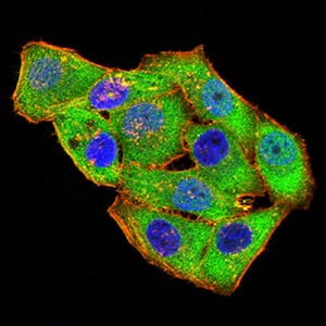
Figure 4:Immunofluorescence analysis of Hela cells using GRIK4 mouse mAb (green). Blue: DRAQ5 fluorescent DNA dye. Red: Actin filaments have been labeled with Alexa Fluor- 555 phalloidin. Secondary antibody from Fisher (Cat#: 35503)
Flow cytometric
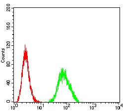
Figure 5:Flow cytometric analysis of SH-SY5Y cells using GRIK4 mouse mAb (green) and negative control (red).
Immunohistochemical analysis
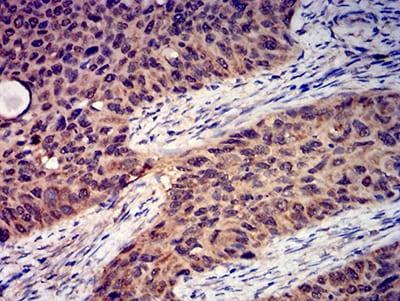
Figure 6:Immunohistochemical analysis of paraffin-embedded cervical cancer tissues using GRIK4 mouse mAb with DAB staining.
Immunohistochemical analysis
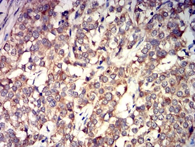
Figure 7:Immunohistochemical analysis of paraffin-embedded bladder cancer tissues using GRIK4 mouse mAb with DAB staining.
For Research Use Only. Not for use in diagnostic procedures.