GRIA2 Primary Antibody
Item Information
Catalog #
Size
Price
Description
Glutamate receptors are the predominant excitatory neurotransmitter receptors in the mammalian brain and are activated in a variety of normal neurophysiologic processes. This gene product belongs to a family of glutamate receptors that are sensitive to alpha-amino-3-hydroxy-5-methyl-4-isoxazole propionate (AMPA), and function as ligand-activated cation channels. These channels are assembled from 4 related subunits, GRIA1-4. The subunit encoded by this gene (GRIA2) is subject to RNA editing (CAG->CGG; Q->R) within the second transmembrane domain, which is thought to render the channel impermeable to Ca(2+). Human and animal studies suggest that pre-mRNA editing is essential for brain function, and defective GRIA2 RNA editing at the Q/R site may be relevant to amyotrophic lateral sclerosis (ALS) etiology. Alternative splicing, resulting in transcript variants encoding different isoforms, (including the flip and flop isoforms that vary in their signal transduction properties), has been noted for this gene.
Product Overview
Entrez GenelD
2891
Aliases
GLUR2; GLURB; GluA2; HBGR2; GluR-K2
Clone#
7A7A3
Host / Isotype
Mouse / IgG2b
Species Reactivity
Human
Immunogen
Purified recombinant fragment of human GRIA2 (AA: 35-175) expressed in E. Coli.
Formulation
Purified antibody in PBS with 0.05% sodium azide
Storage
Store at 4°C short term. Aliquot and store at -20°C long term. Avoid freeze/thaw cycles.
Product Applications
WB (Western Blot)
1/500 - 1/2000
IHC_P(Immunohistochemistry)
1/200 - 1/1000
FCM (Flow Cytometry)
1/200 - 1/400
ELISA
1/10000
References
1.Histopathology. 2014 Jul;65(1):71-80.
2.Proc Natl Acad Sci U S A. 2011 Jan 4;108(1):367-72.
2.Proc Natl Acad Sci U S A. 2011 Jan 4;108(1):367-72.
Product Image
Elisa

Figure 1: Black line: Control Antigen (100 ng);Purple line: Antigen (10ng); Blue line: Antigen (50 ng); Red line:Antigen (100 ng)
Western Blot

Figure 2:Western blot analysis using GRIA2 mAb against human GRIA2 (AA: 35-175) recombinant protein. (Expected MW is 41.9 kDa)
Western Blot

Figure 3:Western blot analysis using GRIA2 mAb against HEK293 (1) and GRIA2 (AA: 35-175)-hIgGFc transfected HEK293 (2) cell lysate.
Flow cytometric

Figure 4:Flow cytometric analysis of SK-N-SH cells using GRIA2 mouse mAb (green) and negative control (red).
Immunohistochemical analysis

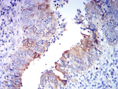
Figure 5:Immunohistochemical analysis of paraffin-embedded rectum cancer tissues using GRIA2 mouse mAb with DAB staining.
For Research Use Only. Not for use in diagnostic procedures.

