

GNAS Primary Antibody
Item Information
Catalog #
Size
Price
Description
This locus has a highly complex imprinted expression pattern. It gives rise to maternally, paternally, and biallelically expressed transcripts that are derived from four alternative promoters and 5' exons. Some transcripts contain a differentially methylated region (DMR) at their 5' exons, and this DMR is commonly found in imprinted genes and correlates with transcript expression. An antisense transcript is produced from an overlapping locus on the opposite strand. One of the transcripts produced from this locus, and the antisense transcript, are paternally expressed noncoding RNAs, and may regulate imprinting in this region. In addition, one of the transcripts contains a second overlapping ORF, which encodes a structurally unrelated protein - Alex. Alternative splicing of downstream exons is also observed, which results in different forms of the stimulatory G-protein alpha subunit, a key element of the classical signal transduction pathway linking receptor-ligand interactions with the activation of adenylyl cyclase and a variety of cellular reponses. Multiple transcript variants encoding different isoforms have been found for this gene. Mutations in this gene result in pseudohypoparathyroidism type 1a, pseudohypoparathyroidism type 1b, Albright hereditary osteodystrophy, pseudopseudohypoparathyroidism, McCune-Albright syndrome, progressive osseus heteroplasia, polyostotic fibrous dysplasia of bone, and some pituitary tumors. [provided by RefSeq, Aug 2012]
Product Overview
Entrez GenelD
2778
Aliases
AHO; GSA; GSP; POH; GPSA; NESP; GNAS1; PHP1A; PHP1B; PHP1C; C20orf45
Clone#
7G6G5
Host / Isotype
Mouse / IgG1
Species Reactivity
Human
Immunogen
Purified recombinant fragment of human GNAS (AA: 42-188) expressed in E. Coli.
Formulation
Purified antibody from tissue culture in PBS with 0.05% sodium azide
Storage
Store at 4°C short term. Aliquot and store at -20°C long term. Avoid freeze/thaw cycles.
Product Applications
WB (Western Blot)
1/500 - 1/2000
ICC (Immunocytochemistry)
1/200 - 1/1000
FCM (Flow Cytometry)
1/200 - 1/400
ELISA
1/10000
References
Br J Cancer. 2013 Mar 5;108(4):951-8.
Anticancer Res. 2012 May;32(5):2169-72.
Anticancer Res. 2012 May;32(5):2169-72.
Product Image
Elisa
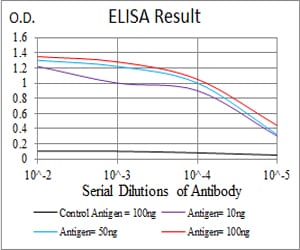
Figure 1: Black line: Control Antigen (100 ng); Purple line: Antigen(10ng); Blue line: Antigen (50 ng); Red line: Antigen (100 ng);
Western Blot
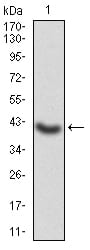
Figure 2:Western blot analysis using GNAS mAb against human GNAS (AA: 42-188) recombinant protein. (Expected MW is 42.8 kDa)
Western Blot
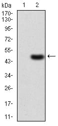
Figure 3:Western blot analysis using GNAS mAb against HEK293 (1) and GNAS (AA: 42-188)-hIgGFc transfected HEK293 (2) cell lysate.
Immunofluorescence analysis
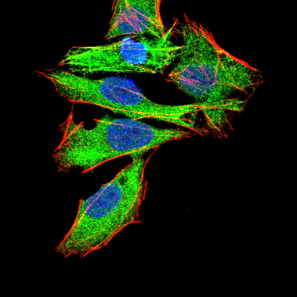
Figure 4:Immunofluorescence analysis of Hela cells using GNAS mouse mAb (green). Blue: DRAQ5 fluorescent DNA dye. Red: Actin filaments have been labeled with Alexa Fluor- 555 phalloidin. Secondary antibody from Fisher (Cat#: 35503)
Flow cytometric
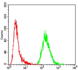
Figure 5:Flow cytometric analysis of MCF-7 cells using GNAS mouse mAb (green) and negative control (red).
For Research Use Only. Not for use in diagnostic procedures.