
GKAP Primary Antibody
Item Information
Catalog #
Size
Price
Description
Function: Part of the postsynaptic scaffold in neuronal cells.Tissue specificity: Expressed in brain.
Product Overview
Entrez GenelD
9229
Aliases
GKAP; DLGAP1; DAP-1; hGKAP; SAPAP1; FLJ38442; MGC88156; DAP-1-BETA; DAP-1-ALPHA
Clone#
3G4
Host / Isotype
Mouse / IgG1
Species Reactivity
Human
Immunogen
Purified recombinant fragment of human GKAP expressed in E. Coli.
Formulation
Ascitic fluid containing 0.03% sodium azide.
Storage
Store at 4°C short term. Aliquot and store at -20°C long term. Avoid freeze/thaw cycles.
Product Applications
WB (Western Blot)
1/500 - 1/2000
IHC_P(Immunohistochemistry)
1/200 - 1/1000
ICC (Immunocytochemistry)
1/200 - 1/1000
ELISA
1/10000
References
1. Am J Hum Genet. 2009 Nov;85(5):628-42.
2. Genes Immun. 2010 Apr;11(3):232-8.
2. Genes Immun. 2010 Apr;11(3):232-8.
Product Image
Western Blot

Figure 1: Western blot analysis using GKAP mAb against HEK293 (1) and GKAP(AA: 490-663)-hIgGFc transfected HEK293 (2) cell lysate.
Immunohistochemical analysis

Figure 2: Immunohistochemical analysis of paraffin-embedded human liver cancer tissues using GKAP mouse mAb with DAB staining.
Immunohistochemical analysis

Figure 3: Immunohistochemical analysis of paraffin-embedded human esophagus tissues using GKAP mouse mAb with DAB staining.
Immunofluorescence analysis
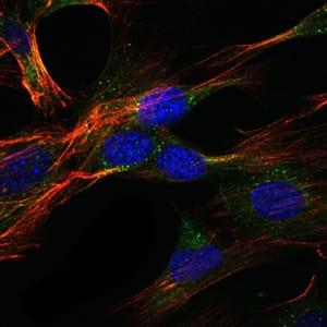
Figure 4: Immunofluorescence analysis of NIH/3T3 cells using GKAP mouse mAb (green). Blue: DRAQ5 fluorescent DNA dye. Red: Actin filaments have been labeled with Alexa Fluor-555 phalloidin.
Elisa

Red: Control Antigen (100ng); Purple: Antigen (10ng); Green: Antigen (50ng); Blue: Antigen (100ng);
For Research Use Only. Not for use in diagnostic procedures.