

GH1 Primary Antibody
Item Information
Catalog #
Size
Price
Description
The protein encoded by this gene is a member of the somatotropin/prolactin family of hormones which play an important role in growth control. The gene, along with four other related genes, is located at the growth hormone locus on chromosome 17 where they are interspersed in the same transcriptional orientation; an arrangement which is thought to have evolved by a series of gene duplications. The five genes share a remarkably high degree of sequence identity. Alternative splicing generates additional isoforms of each of the five growth hormones, leading to further diversity and potential for specialization. This particular family member is expressed in the pituitary but not in placental tissue as is the case for the other four genes in the growth hormone locus. Mutations in or deletions of the gene lead to growth hormone deficiency and short stature.
Product Overview
Entrez GenelD
2688
Aliases
GH; GHN; GH-N; GHB5; hGH-N; IGHD1B
Clone#
3H1C2
Host / Isotype
Mouse / IgG1
Species Reactivity
Human
Immunogen
Purified recombinant fragment of human GH1 (AA: 1-217) expressed in E. Coli.
Formulation
Purified antibody in PBS with 0.05% sodium azide
Storage
Store at 4°C short term. Aliquot and store at -20°C long term. Avoid freeze/thaw cycles.
Product Applications
WB (Western Blot)
1/500 - 1/2000
FCM (Flow Cytometry)
1/200 - 1/400
ELISA
1/10000
References
1.Asian Pac J Cancer Prev. 2015;16(13):5421-5.
2.Tumour Biol. 2014 May;35(5):4529-38.
2.Tumour Biol. 2014 May;35(5):4529-38.
Product Image
Elisa
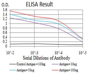
Figure 1: Black line: Control Antigen (100 ng);Purple line: Antigen (10ng); Blue line: Antigen (50 ng); Red line:Antigen (100 ng)
Western Blot
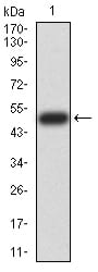
Figure 2:Western blot analysis using GH1 mAb against human GH1 (AA: 1-217) recombinant protein. (Expected MW is 50.8 kDa)
Western Blot
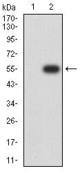
Figure 3:Western blot analysis using GH1 mAb against HEK293 (1) and GH1 (AA: 1-217)-hIgGFc transfected HEK293 (2) cell lysate.
Flow cytometric
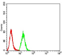
Figure 4:Flow cytometric analysis of Hela cells using GH1 mouse mAb (green) and negative control (red).
For Research Use Only. Not for use in diagnostic procedures.