

GFPT1 Primary Antibody
Item Information
Catalog #
Size
Price
Description
This gene encodes the first and rate-limiting enzyme of the hexosamine pathway and controls the flux of glucose into the hexosamine pathway. The product of this gene catalyzes the formation of glucosamine 6-phosphate.
Product Overview
Entrez GenelD
2673
Aliases
GFA; GFAT; GFPT; MSLG; CMS12; GFAT1; CMSTA1; GFAT 1; GFAT1m; GFPT1L
Clone#
1F1B9
Host / Isotype
Mouse / IgG1
Species Reactivity
Human, Monkey, Rat
Immunogen
Purified recombinant fragment of human GFPT1 (AA: 536-681) expressed in E. Coli.
Formulation
Purified antibody in PBS with 0.05% sodium azide
Storage
Store at 4°C short term. Aliquot and store at -20°C long term. Avoid freeze/thaw cycles.
Product Applications
WB (Western Blot)
1/500 - 1/2000
ICC (Immunocytochemistry)
1/200 - 1/1000
FCM (Flow Cytometry)
1/200 - 1/400
ELISA
1/10000
References
1.Neurology. 2013 Jul 23;81(4):370-8.
2.Hum Mol Genet. 2013 Jul 15;22(14):2905-13.
2.Hum Mol Genet. 2013 Jul 15;22(14):2905-13.
Product Image
Elisa
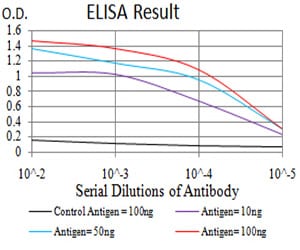
Figure 1: Black line: Control Antigen (100 ng);Purple line: Antigen (10ng); Blue line: Antigen (50 ng); Red line:Antigen (100 ng)
Western Blot

Figure 2:Western blot analysis using GFPT1 mAb against human GFPT1 (AA: 536-681) recombinant protein. (Expected MW is 42.3 kDa)
Western Blot
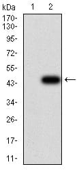
Figure 3:Western blot analysis using GFPT1 mAb against HEK293 (1) and GFPT1 (AA: 536-681)-hIgGFc transfected HEK293 (2) cell lysate.
Western Blot

Figure 4:Western blot analysis using GFPT1 mouse mAb against Hela (1), HepG2 (2), HEK293 (3), BEL-7402 (4), SMMC-7721 (5), SK-MES-1 (6), C6 (7), and COS7 (8) cell lysate.
Immunofluorescence analysis

Figure 5:Immunofluorescence analysis of Hela cells using GFPT1 mouse mAb (green). Blue: DRAQ5 fluorescent DNA dye. Red: Actin filaments have been labeled with Alexa Fluor- 555 phalloidin. Secondary antibody from Fisher (Cat#: 35503)
Flow cytometric

Figure 6:Flow cytometric analysis of Hela cells using GFPT1 mouse mAb (green) and negative control (red).
For Research Use Only. Not for use in diagnostic procedures.