
G6PD Primary Antibody
Item Information
Catalog #
Size
Price
Description
This gene encodes glucose-6-phosphate dehydrogenase. This protein is a cytosolic enzyme encoded by a housekeeping X-linked gene whose main function is to produce NADPH, a key electron donor in the defense against oxidizing agents and in reductive biosynthetic reactions. G6PD is remarkable for its genetic diversity. Many variants of G6PD, mostly produced from missense mutations, have been described with wide ranging levels of enzyme activity and associated clinical symptoms. G6PD deficiency may cause neonatal jaundice, acute hemolysis, or severe chronic non-spherocytic hemolytic anemia. Two transcript variants encoding different isoforms have been found for this gene.
Product Overview
Entrez GenelD
2539
Aliases
G6PD1
Clone#
5E12
Host / Isotype
Mouse / IgG1
Species Reactivity
Human
Immunogen
Purified recombinant fragment of human G6PD expressed in E. Coli.
Formulation
Ascitic fluid containing 0.03% sodium azide.
Storage
Store at 4°C short term. Aliquot and store at -20°C long term. Avoid freeze/thaw cycles.
Product Applications
WB (Western Blot)
1/500 - 1/2000
IHC_P(Immunohistochemistry)
1/200 - 1/1000
FCM (Flow Cytometry)
1/200 - 1/400
ELISA
1/10000
References
1. Science. 2009 Dec 11;326(5959):1546-9.
2. Immunol Invest. 2009;38(6):551-9.
2. Immunol Invest. 2009;38(6):551-9.
Product Image
Western Blot

Figure 1: Western blot analysis using G6PD mAb against human G6PD (AA: 275-515) recombinant protein.(Expected MW is 53.1 kDa)
Western Blot

Figure 2: Western blot analysis using G6PD mouse mAb against Hela (1), MCF-7 (2), Jurkat (3) and K562 (4) cell lysate.
Immunohistochemical analysis

Figure 3: Immunohistochemical analysis of paraffin-embedded ovarian cancer tissues using G6PD mouse mAb with DAB staining.
Immunohistochemical analysis
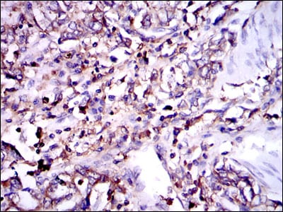
Figure 4: Immunohistochemical analysis of paraffin-embedded stomach cancer tissues using G6PD mouse mAb with DAB staining.
Flow cytometric

Figure 5: Flow cytometric analysis of MCF-7 cells using G6PD mouse mAb (green) and negative control (red).
Elisa

Black line: Control Antigen (100 ng); Purple line: Antigen(10ng); Blue line: Antigen (50 ng); Red line: Antigen (100 ng);
For Research Use Only. Not for use in diagnostic procedures.