

FUT4 Primary Antibody
Item Information
Catalog #
Size
Price
Description
The product of this gene transfers fucose to N-acetyllactosamine polysaccharides to generate fucosylated carbohydrate structures. It catalyzes the synthesis of the non-sialylated antigen, Lewis x (CD15).
Product Overview
Entrez GenelD
2526
Aliases
LeX; CD15; ELFT; FCT3A; FUTIV; SSEA-1; FUC-TIV
Clone#
6B11B4
Host / Isotype
Mouse / IgG2b
Species Reactivity
Human
Immunogen
Purified recombinant fragment of human FUT4 (AA: 199-302) expressed in E. Coli.
Formulation
Purified antibody in PBS with 0.05% sodium azide
Storage
Store at 4°C short term. Aliquot and store at -20°C long term. Avoid freeze/thaw cycles.
Product Applications
WB (Western Blot)
1/500 - 1/2000
ELISA
1/10000
References
1. J Immunol. 2011 Dec 15;187(12):6227-34.
2. PLoS One. 2011;6(9):e24584.
2. PLoS One. 2011;6(9):e24584.
Product Image
Western Blot

Figure 1: Western blot analysis using FUT4 mAb against human FUT4 recombinant protein. (Expected MW is 37 kDa)
Western Blot

Figure 2: Western blot analysis using FUT4 mAb against HEK293 (1) and FUT4 (AA: 199-302)-hIgGFc transfected HEK293 (2) cell lysate.
Western Blot
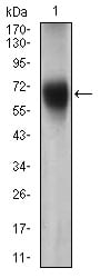
Figure 3: Western blot analysis using FUT4 mouse mAb against Jurkat cell lysate.
Elisa

Black line: Control Antigen (100 ng); Purple line: Antigen(10ng); Blue line: Antigen (50 ng); Red line: Antigen (100 ng);
For Research Use Only. Not for use in diagnostic procedures.