

FSHR Primary Antibody
Item Information
Catalog #
Size
Price
Description
The protein encoded by this gene belongs to family 1 of G-protein coupled receptors. It is the receptor for follicle stimulating hormone and functions in gonad development. Mutations in this gene cause ovarian dysgenesis type 1, and also ovarian hyperstimulation syndrome. Alternative splicing results in multiple transcript variants.
Product Overview
Entrez GenelD
2492
Aliases
LGR1; ODG1; FSHR1; FSHRO
Clone#
3D5G9
Host / Isotype
Mouse / Mouse IgG1
Immunogen
Purified recombinant fragment of human FSHR (AA: extra 18-366) expressed in E. Coli.
Formulation
Purified antibody in PBS with 0.05% sodium azide
Storage
Store at 4°C short term. Aliquot and store at -20°C long term. Avoid freeze/thaw cycles.
Product Applications
WB (Western Blot)
1/500 - 1/2000
FCM (Flow Cytometry)
1/200 - 1/400
ELISA
1/10000
References
1.Endocrinology. 2018 Aug 1;159(8):3020-3035. 2.Endokrynol Pol. 2018;69(2):192-198.
Product Image
Elisa

Figure 1:Black line: Control Antigen (100 ng);Purple line: Antigen (10ng); Blue line: Antigen (50 ng); Red line:Antigen (100 ng)
Western Blot

Figure 2:Western blot analysis using FSHR mAb against human FSHR (AA: 18-366) recombinant protein. (Expected MW is 42.8 kDa)
Western Blot
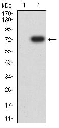
Figure 3:Western blot analysis using FSHR mAb against HEK293 (1) and FSHR (AA: 18-366)-hIgGFc transfected HEK293 (2) cell lysate.
Flow Cytometric

Figure 4:Flow cytometric analysis of Hela cells using FSHR mouse mAb (green) and negative control (red).
For Research Use Only. Not for use in diagnostic procedures.