

FSHB Primary Antibody
Item Information
Catalog #
Size
Price
Description
The pituitary glycoprotein hormone family includes follicle-stimulating hormone, luteinizing hormone, chorionic gonadotropin, and thyroid-stimulating hormone. All of these glycoproteins consist of an identical alpha subunit and a hormone-specific beta subunit. This gene encodes the beta subunit of follicle-stimulating hormone. In conjunction with luteinizing hormone, follicle-stimulating hormone induces egg and sperm production. Alternative splicing results in two transcript variants encoding the same protein.
Product Overview
Entrez GenelD
2488
Aliases
HH24
Clone#
2H7B1
Host / Isotype
Mouse / Mouse IgG2a
Species Reactivity
Human
Immunogen
Purified recombinant fragment of human FSHB (AA: 2x 19-129) expressed in E. Coli.
Formulation
Purified antibody in PBS with 0.05% sodium azide
Storage
Store at 4°C short term. Aliquot and store at -20°C long term. Avoid freeze/thaw cycles.
Product Applications
WB (Western Blot)
1/500 - 1/2000
IHC_P(Immunohistochemistry)
1/200 - 1/1000
FCM (Flow Cytometry)
1/200 - 1/400
ELISA
1/10000
References
1.BMC Endocr Disord. 2018 Feb 7;18(1):8.
2.J Clin Endocrinol Metab. 2016 May;101(5):2178-84.
2.J Clin Endocrinol Metab. 2016 May;101(5):2178-84.
Product Image
Elisa

Figure 1:Black line: Control Antigen (100 ng);Purple line: Antigen (10ng); Blue line: Antigen (50 ng); Red line:Antigen (100 ng)
Western Blot
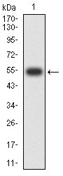
Figure 2:Western blot analysis using FSHB mAb against human FSHB (AA: 2x 19-129) recombinant protein. (Expected MW is 54.1 kDa)
Western Blot
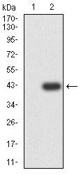
Figure 3:Western blot analysis using FSHB mAb against HEK293-6e (1) and FSHB (AA: 2x 19-129)-hIgGFc transfected HEK293-6e (2) cell lysate.
Immunofluorescence analysis
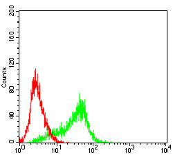
Figure 4:Flow cytometric analysis of Hela cells using FSHB mouse mAb (green) and negative control (red).
Immunohistochemical analysis

Figure 5:Immunohistochemical analysis of paraffin-embedded human hypophysis tissues using FSHB mouse mAb with DAB staining.
For Research Use Only. Not for use in diagnostic procedures.