

FOXP2 Primary Antibody
Item Information
Catalog #
Size
Price
Description
This gene encodes a member of the forkhead/winged-helix (FOX) family of transcription factors. It is expressed in fetal and adult brain as well as in several other organs such as the lung and gut. The protein product contains a FOX DNA-binding domain and a large polyglutamine tract and is an evolutionarily conserved transcription factor, which may bind directly to approximately 300 to 400 gene promoters in the human genome to regulate the expression of a variety of genes. This gene is required for proper development of speech and language regions of the brain during embryogenesis, and may be involved in a variety of biological pathways and cascades that may ultimately influence language development. Mutations in this gene cause speech-language disorder 1 (SPCH1), also known as autosomal dominant speech and language disorder with orofacial dyspraxia. Multiple alternative transcripts encoding different isoforms have been identified in this gene.
Product Overview
Entrez GenelD
93986
Aliases
SPCH1; CAGH44; TNRC10
Clone#
2G11B8
Host / Isotype
Mouse / IgG1
Species Reactivity
Human
Immunogen
Purified recombinant fragment of human FOXP2 (AA: 641-740) expressed in E. Coli.
Formulation
Purified antibody in PBS with 0.05% sodium azide
Storage
Store at 4°C short term. Aliquot and store at -20°C long term. Avoid freeze/thaw cycles.
Product Applications
WB (Western Blot)
1/500 - 1/2000
FCM (Flow Cytometry)
1/200 - 1/400
ELISA
1/10000
References
1.J Clin Pathol. 2013 Jul;66(7):563-8.
2.World J Biol Psychiatry. 2013 Mar;14(2):146-50.
2.World J Biol Psychiatry. 2013 Mar;14(2):146-50.
Product Image
Elisa

Figure 1: Black line: Control Antigen (100 ng);Purple line: Antigen (10ng); Blue line: Antigen (50 ng); Red line:Antigen (100 ng)
Western Blot

Figure 2:Western blot analysis using FOXP2 mAb against human FOXP2 (AA: 641-740) recombinant protein. (Expected MW is 36.7 kDa)
Western Blot
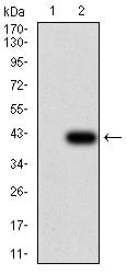
Figure 3:Western blot analysis using FOXP2 mAb against HEK293 (1) and FOXP2 (AA: 641-740)-hIgGFc transfected HEK293 (2) cell lysate.
Western Blot

Figure 4:Western blot analysis using FOXP2 mouse mAb against HepG2 (1) cell lysate.
Flow cytometric

Figure 5:Flow cytometric analysis of HeLa cells using FOXP2 mouse mAb (green) and negative control (red).
For Research Use Only. Not for use in diagnostic procedures.