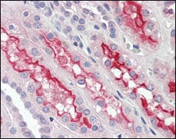
FER Primary Antibody

Item Information
Catalog #
Size
Price
Description

FER (fer tyrosine kinase) is a member of the FPS/FES family of nontransmembrane receptor tyrosine kinases, which shares a functional domain and is involved in signaling pathways through receptor tyrosine kinases (RTK) and cytokine receptors. The Fes /Fps family is distinct from c-Src, c-Abl and related nRTKs and was originally distinguished as a homolog to retroviral oncoproteins. In vivo, Fer kinase assembles into homotrimers via conserved coiled-coil domains. The N-terminal coiled-coil domains of Fer can autophosphorylate in trans, thereby regulating their cellular function through differential phosphorylation states. Growth factor exposure can induce tyrosine phosphorylation of Fer and recruitment of Fer to RTK complexes containing p85. It is expressed predominantly in mature hematopoietic cells of the granulocytic and monocytic lineage, and has been shown to be expressed in vascular endothelial cells. Fer is implicated in insulin signaling, cell-cell signaling, human prostatic proliferative diseases, and is involved in the regulation of G1 progression.

Product Overview
Entrez GenelD
2241
Aliases
TYK3; FER
Clone#
5D2C4
Host / Isotype
Mouse / IgG1
Species Reactivity
Human, Mouse
Immunogen
Purified recombinant fragment of human FER expressed in E. Coli.
Formulation
Ascitic fluid containing 0.03% sodium azide.
Storage
4°C; -20°C for long term storage
Product Applications
WB (Western Blot)
1/500 - 1/2000
IHC_P(Immunohistochemistry)
1/200 - 1/1000
ICC (Immunocytochemistry)
1/200 - 1/1000
ELISA
1/10000
References
1. Li L. Cheng X. Ling HQ. Plant Mol Biol. 2004, Jan, 54(1):125-36.
2. Girault JA. Greengard P. Arch Neurol. 2004, May, 61(5):641-4.
3. Fan L. Di Ciano-Oliveira C. Weed SA. et al. Biochem J. 2004, Jun 1, 380(Pt 2):581-91.
Product Image
Western Blot
Figure 1: Western blot analysis using FER mouse mAb against NIH/3T3 (1), A549 (2) and SK-MEL-5 (3) cell lysate.
Immunohistochemical analysis
Figure 2: Immunohistochemical analysis of paraffin-embedded human kidney tissues using FER mouse mAb.
Immunofluorescence analysis
Figure 3: Confocal Immunofluorescence analysis of Hela cells using FER mouse mAb (green). Red: Actin filaments have been labeled with Alexa Fluor-555 phalloidin. Blue: DRAQ5 fluorescent DNA dye.
For Research Use Only. Not for use in diagnostic procedures.