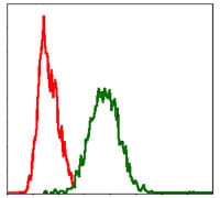
FAS Primary Antibody

Item Information
Catalog #
Size
Price
Description

The protein encoded by this gene is a member of the TNF-receptor superfamily. This receptor contains a death domain. It has been shown to play a central role in the physiological regulation of programmed cell death, and has been implicated in the pathogenesis of various malignancies and diseases of the immune system. The interaction of this receptor with its ligand allows the formation of a death-inducing signaling complex that includes Fas-associated death domain protein (FADD), caspase 8, and caspase 10. The autoproteolytic processing of the caspases in the complex triggers a downstream caspase cascade, and leads to apoptosis. This receptor has been also shown to activate NF-kappaB, MAPK3/ERK1, and MAPK8/JNK, and is found to be involved in transducing the proliferating signals in normal diploid fibroblast and T cells. Several alternatively spliced transcript variants have been described, some of which are candidates for nonsense-mediated mRNA decay (NMD). The isoforms lacking the transmembrane domain may negatively regulate the apoptosis mediated by the full length isoform.

Product Overview
Entrez GenelD
355
Aliases
APT1; CD95; FAS1; APO-1; FASTM; ALPS1A; TNFRSF6
Clone#
4F8H6
Host / Isotype
Mouse / IgG1
Species Reactivity
Human
Immunogen
Purified recombinant fragment of human FAS expressed in E. Coli.
Formulation
Purified antibody in PBS with 0.05% sodium azide
Storage
4°C; -20°C for long term storage
Product Applications
WB (Western Blot)
1/500 - 1/2000
ICC (Immunocytochemistry)
1/200 - 1/1000
FCM (Flow Cytometry)
1/200 - 1/400
ELISA
1/10000
References
Am J Hum Genet. 2009 Nov;85(5):628-42
Int J Surg Pathol. 2010 Dec;18(6):493-8
Product Image
Elisa
Figure 1: Black line: Control Antigen (100 ng); Purple line: Antigen(10ng); Blue line: Antigen (50 ng); Red line: Antigen (100 ng);
Western Blot
Figure 2: Western blot analysis using FAS mAb against human FAS (AA: 87-278) recombinant protein. (Expected MW is 47.2 kDa)
Western Blot
Figure 3: Western blot analysis using FAS mouse mAb against Hela (1), Jurkat (2) cell lysate.
Immunofluorescence analysis
Figure 4: Immunofluorescence analysis of Hela cells using FAS mouse mAb (green). Blue: DRAQ5 fluorescent DNA dye. Red: Actin filaments have been labeled with Alexa Fluor-555 phalloidin.
Flow cytometric
Figure 5: Flow cytometric analysis of Hela cells using FAS mouse mAb (green) and negative control (red).
For Research Use Only. Not for use in diagnostic procedures.