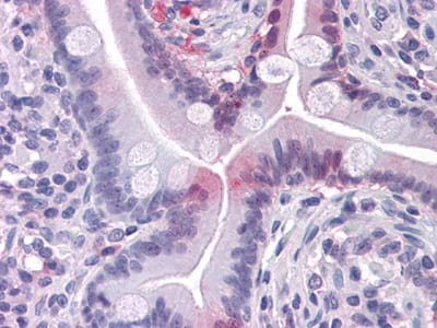
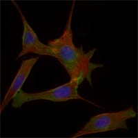
FABP2 Primary Antibody

Item Information
Catalog #
Size
Price
Description

The intracellular fatty acid-binding proteins (FABPs) belong to a multigene family with nearly twenty identified members. FABPs are divided into at least three distinct types, namely the hepatic-, intestinal- and cardiac-type. They form 14-15 kDa proteins and are thought to participate in the uptake, intracellular metabolism and/or transport of long-chain fatty acids. They may also be responsible in the modulation of cell growth and proliferation. Intestinal fatty acid-binding protein 2 gene contains four exons and is an abundant cytosolic protein in small intestine epithelial cells. This gene has a polymorphism at codon 54 that identified an alanine-encoding allele and a threonine-encoding allele. Thr-54 protein is associated with increased fat oxidation and insulin resistance. Genetic variation in FABP2 may thus contribute to interindividual variation in the response of plasma lipoproteins to different dietary fibres, but the mechanism does not appear to be related to increases in fecal bile acid secretion.

Product Overview
Entrez GenelD
2169
Aliases
FABPI; I-FABP; MGC133132; FABP2
Clone#
9A9B7B3
Host / Isotype
Mouse / IgG1
Species Reactivity
Human
Immunogen
Purified recombinant fragment of human FABP2 expressed in E. Coli.
Formulation
Ascitic fluid containing 0.03% sodium azide.
Storage
4°C; -20°C for long term storage
Product Applications
WB (Western Blot)
1/500 - 1/2000
IHC_P(Immunohistochemistry)
1/200 - 1/1000
ICC (Immunocytochemistry)
1/200 - 1/1000
FCM (Flow Cytometry)
1/200 - 1/400
ELISA
1/10000
References
1. Yamada, K. et al. (1997) Diabetologia. 40(6):706-10
2. Georgopoulos, A. et al. (2000)85(9):3155-60
3. Kim, CH. et al. (2001) Metabolism. 50(4):473-6
4. Fisher, E. et al. (2006) Horm Metab Res. 38(5):341-5
Product Image
Western Blot
Figure 1: Western blot analysis using FABP2 mouse mAb against FABP2-hIgGFc transfected HEK293 (1) cell lysate and LOVO (2) cell lysate.
Immunohistochemical analysis
Figure 2: Immunohistochemical analysis of paraffin-embedded human Small Intestine tissues using FABP2 mouse mAb
Flow cytometric
Figure 3: Flow cytometric analysis of LOVO cells using FABP2 mouse mAb (green) and negative control (purple).
Immunofluorescence analysis
Figure 3: Immunofluorescence analysis of 3T3-L1 cells using FABP2 mouse mAb (green). Blue: DRAQ5 fluorescent DNA dye. Red: Actin filaments have been labeled with Alexa Fluor-555 phalloidin.
For Research Use Only. Not for use in diagnostic procedures.