

EZR Primary Antibody
Item Information
Catalog #
Size
Price
Description
The cytoplasmic peripheral membrane protein encoded by this gene functions as a protein-tyrosine kinase substrate in microvilli. As a member of the ERM protein family, this protein serves as an intermediate between the plasma membrane and the actin cytoskeleton. This protein plays a key role in cell surface structure adhesion, migration and organization, and it has been implicated in various human cancers. A pseudogene located on chromosome 3 has been identified for this gene. Alternatively spliced variants have also been described for this gene.
Product Overview
Entrez GenelD
7430
Aliases
CVL; CVIL; VIL2; HEL-S-105
Clone#
6F1A9
Host / Isotype
Mouse / IgG1
Species Reactivity
Human, Mouse, Monkey
Immunogen
Purified recombinant fragment of human EZR (AA: 292-464) expressed in E. Coli.
Formulation
Purified antibody from tissue culture in PBS with 0.05% sodium azide
Storage
Store at 4°C short term. Aliquot and store at -20°C long term. Avoid freeze/thaw cycles.
Product Applications
WB (Western Blot)
1/500 - 1/2000
ICC (Immunocytochemistry)
1/100 - 1/400
FCM (Flow Cytometry)
1/200 - 1/400
ELISA
1/10000
References
1.BMC Cancer. 2013 Nov 4;13:520.
2.Cell Oncol (Dordr). 2013 Dec;36(6):485-91.
2.Cell Oncol (Dordr). 2013 Dec;36(6):485-91.
Product Image
Elisa
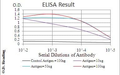
Figure 1: Black line: Control Antigen (100 ng); Purple line: Antigen(10ng); Blue line: Antigen (50 ng); Red line: Antigen (100 ng);
Western Blot
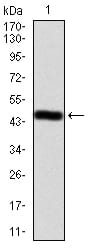
Figure 2:Western blot analysis using EZR mAb against human EZR (AA: 292-464) recombinant protein. (Expected MW is 47.1 kDa)
Western Blot
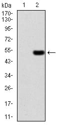
Figure 3:Western blot analysis using EZR mAb against HEK293 (1) and EZR (AA: 292-464)-hIgGFc transfected HEK293 (2) cell lysate.
Western Blot
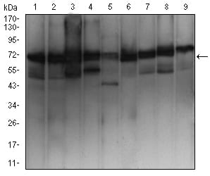
Figure 4:Western blot analysis using EZR mouse mAb against MCF-7 (1), Hela (2), A431 (3), Hek293 (4), SK-N-SH (5), Jurkat (6), HepG2 (7), NIH/3T3 (8), and Cos7 (9) cell lysate.
Immunofluorescence analysis
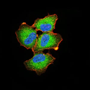
Figure 5:Immunofluorescence analysis of A549 cells using EZR mouse mAb (green). Blue: DRAQ5 fluorescent DNA dye. Red: Actin filaments have been labeled with Alexa Fluor- 555 phalloidin. Secondary antibody from Fisher (Cat#: 35503)
Flow cytometric
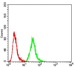
Figure 6:Flow cytometric analysis of MCF-7 cells using EZR mouse mAb (green) and negative control (red).
Flow cytometric

Figure 7:Flow cytometric analysis of Hela cells using EZR mouse mAb (green) and negative control (red).
For Research Use Only. Not for use in diagnostic procedures.