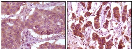
ERK2 Primary Antibody

Item Information
Catalog #
Size
Price
Description

ERK2 (also designated extracellular-signal-related kinase 2 or mitogen-activated protein kinase 1), with 360-amino acid protein (about 40kDa), belongs to the MAP kinase family. MAP kinases act as an integration point for multiple biochemical signals, and are involved in a wide variety of cellular processes such as proliferation, differentiation, transcription regulation and development. The activation of ERK2 requires its phosphorylation by upstream kinases. ERK2 is located in the cytoplasm of resting cells and translocates into the nucleus upon extracellular stimuli by active transport of a dimer. ERK2 is essential for placental development and ERK2 in the trophoblast compartment may be indispensable for the vascularization of the labyrinth.

Product Overview
Entrez GenelD
5594
Aliases
ERK; p38; p40; p41; ERT1; MAPK2; PRKM1; P42MAPK; p41mapk; MAPK1
Clone#
4C11
Host / Isotype
Mouse / IgG2a
Species Reactivity
Human, Mouse, Monkey
Immunogen
Purified recombinant fragment of human ERK2 expressed in E. Coli.
Formulation
Purified antibody in PBS containing 0.03% sodium azide.
Storage
4°C; -20°C for long term storage
Product Applications
WB (Western Blot)
1/500 - 1/2000
IHC_P(Immunohistochemistry)
1/200 - 1/1000
ICC (Immunocytochemistry)
1/200 - 1/1000
ELISA
1/10000
References
1. Angelique W. Whitehurst, Fred L. Robinson, Mary Shannon Moore. J. Biol. Chem., Mar 2004; 279: 12840 - 12847.
2. N Hatano, Y Mori, M Oh-hora. Genes Cells, Nov 2003; 8: 847 - 856.
Product Image
Western Blot
Figure 1: Western blot analysis using ERK2 mouse mAb against Hela (1), NIH/3T3 (2), MCF-7 (3), HEK293 (4), Jurkat (5), A549 (6), NTERA-2 (7) and SMMC-7721 (8) cell lysate.
Immunohistochemical analysis
Figure 2: Immunohistochemical analysis of paraffin-embedded human lung carcinoma (left) and breast carcinoma (right) showing cytoplasmic localization using ERK2 mouse mAb with DAB staining.
Immunofluorescence analysis
Figure 3: Confocal Immunofluorescence analysis of Eca-109 cells using ERK2 mouse mAb (green).
For Research Use Only. Not for use in diagnostic procedures.