

ERCC1 Primary Antibody
Item Information
Catalog #
Size
Price
Description
The product of this gene functions in the nucleotide excision repair pathway, and is required for the repair of DNA lesions such as those induced by UV light or formed by electrophilic compounds including cisplatin. The encoded protein forms a heterodimer with the XPF endonuclease (also known as ERCC4), and the heterodimeric endonuclease catalyzes the 5' incision in the process of excising the DNA lesion. The heterodimeric endonuclease is also involved in recombinational DNA repair and in the repair of inter-strand crosslinks. Mutations in this gene result in cerebrooculofacioskeletal syndrome, and polymorphisms that alter expression of this gene may play a role in carcinogenesis. Multiple transcript variants encoding different isoforms have been found for this gene. The last exon of this gene overlaps with the CD3e molecule, epsilon associated protein gene on the opposite strand.
Product Overview
Entrez GenelD
2067
Aliases
UV20; COFS4; RAD10
Clone#
1A5A2
Host / Isotype
Mouse / IgG1
Species Reactivity
Human
Immunogen
Purified recombinant fragment of human ERCC1 (AA: 151-297) expressed in E. Coli.
Formulation
Purified antibody in PBS with 0.05% sodium azide
Storage
Store at 4°C short term. Aliquot and store at -20°C long term. Avoid freeze/thaw cycles.
Product Applications
WB (Western Blot)
1/500 - 1/2000
FCM (Flow Cytometry)
1/200 - 1/400
ELISA
1/10000
References
1.Tumour Biol. 2014 Sep;35(9):8721-31.
2.Anticancer Res. 2014 Jan;34(1):401-6.
2.Anticancer Res. 2014 Jan;34(1):401-6.
Product Image
Elisa
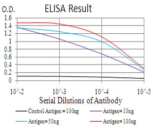
Figure 1: Black line: Control Antigen (100 ng); Purple line: Antigen(10ng); Blue line: Antigen (50 ng); Red line: Antigen (100 ng);
Western Blot
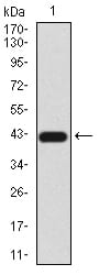
Figure 2:Western blot analysis using ERCC1 mAb against human ERCC1 (AA: 151-297) recombinant protein. (Expected MW is 42.5 kDa)
Western Blot
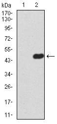
Figure 3:Western blot analysis using ERCC1 mAb against HEK293 (1) and ERCC1 (AA: 151-297)-hIgGFc transfected HEK293 (2) cell lysate.
Flow cytometric

Figure 4:Flow cytometric analysis of Hela cells using ERCC1 mouse mAb (green) and negative control (red).
For Research Use Only. Not for use in diagnostic procedures.