

ERBB4 Primary Antibody
Item Information
Catalog #
Size
Price
Description
This gene is a member of the Tyr protein kinase family and the epidermal growth factor receptor subfamily. It encodes a single-pass type I membrane protein with multiple cysteine rich domains, a transmembrane domain, a tyrosine kinase domain, a phosphotidylinositol-3 kinase binding site and a PDZ domain binding motif. The protein binds to and is activated by neuregulins and other factors and induces a variety of cellular responses including mitogenesis and differentiation. Multiple proteolytic events allow for the release of a cytoplasmic fragment and an extracellular fragment. Mutations in this gene have been associated with cancer. Alternatively spliced variants which encode different protein isoforms have been described; however, not all variants have been fully characterized.
Product Overview
Entrez GenelD
2066
Aliases
HER4; ALS19; p180erbB4
Clone#
4E7G5
Host / Isotype
Mouse / IgG1
Species Reactivity
Human
Immunogen
Purified recombinant fragment of human ERBB4 (AA: 1159-1308) expressed in E. Coli.
Formulation
Purified antibody in PBS with 0.05% sodium azide
Storage
Store at 4°C short term. Aliquot and store at -20°C long term. Avoid freeze/thaw cycles.
Product Applications
WB (Western Blot)
1/500 - 1/2000
ICC (Immunocytochemistry)
1/50 - 1/200
FCM (Flow Cytometry)
1/200 - 1/400
ELISA
1/10000
References
1.Neurosci Lett. 2012 Dec 7;531(2):209-14.
2.EMBO Mol Med. 2013 Jul;5(7):1019-34.
2.EMBO Mol Med. 2013 Jul;5(7):1019-34.
Product Image
Elisa

Figure 1: Black line: Control Antigen (100 ng); Purple line: Antigen(10ng); Blue line: Antigen (50 ng); Red line: Antigen (100 ng);
Western Blot

Figure 2:Western blot analysis using ERBB4 mAb against human ERBB4 (AA: 1159-1308) recombinant protein. (Expected MW is 43.3 kDa)
Western Blot

Figure 3:Western blot analysis using ERBB4 mAb against HEK293 (1) and ERBB4 (AA: 1159-1308)-hIgGFc transfected HEK293 (2) cell lysate.
Immunofluorescence analysis

Figure 4:Immunofluorescence analysis of Hela cells using ERBB4 mouse mAb (green). Blue: DRAQ5 fluorescent DNA dye. Red: Actin filaments have been labeled with Alexa Fluor- 555 phalloidin. Secondary antibody from Fisher (Cat#: 35503)
Flow cytometric
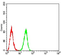
Figure 5:Flow cytometric analysis of Hela cells using ERBB4 mouse mAb (green) and negative control (red).
For Research Use Only. Not for use in diagnostic procedures.