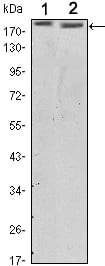
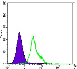
ERBB2 Primary Antibody

Item Information
Catalog #
Size
Price
Description

ERBB2: v-erb-b2 erythroblastic leukemia viral oncogene homolog 2, neuro/glioblastoma derived oncogene homolog (avian). This gene encodes a member of the epidermal growth factor (EGF) receptor family of receptor tyrosine kinases. This protein has no ligand binding domain of its own and therefore cannot bind growth factors. However, it does bind tightly to other ligand-bound EGF receptor family members to form a heterodimer, stabilizing ligand binding and enhancing kinase-mediated activation of downstream signalling pathways, such as those involving mitogen-activated protein kinase and phosphatidylinositol-3 kinase. Allelic variations at amino acid positions 654 and 655 of isoform a (positions 624 and 625 of isoform b) have been reported, with the most common allele, Ile654/Ile655, shown here. Amplification and/or overexpression of this gene has been reported in numerous cancers, including breast and ovarian tumors. Alternative splicing results in several additional transcript variants, some encoding different isoforms and others that have not been fully characterized.

Product Overview
Entrez GenelD
2064
Aliases
NEU; HER2; TKR1; CD340; HER-2
Clone#
6C2
Host / Isotype
Mouse / IgG1
Species Reactivity
Human
Immunogen
Purified recombinant fragment of human ERBB2(aa750-987) expressed in E. Coli.
Formulation
Ascitic fluid containing 0.03% sodium azide.
Storage
4°C; -20°C for long term storage
Product Applications
WB (Western Blot)
1/500 - 1/2000
IHC_P(Immunohistochemistry)
1/200 - 1/1000
FCM (Flow Cytometry)
1/200 - 1/400
ELISA
1/10000
References
1. RR Mehta, JH McDermott, TJ Hieken, et al. J. Clin. Oncol. 1998;16:2409 - 2416.
2. Hideko Y, Vered S, and Daniel F.H, et al. J. Clin. Oncol.2001;19:2334 - 2356.
3. Magali F, Kamel H, Cile B, et al. Clinical Cancer Research. 2000;6:4745-4754.
Product Image
Western Blot
Figure 1: Western blot analysis using ERBB2 mouse mAb against SKBR3 (1) and MCF-7 (2) cell lysate.
Immunohistochemical analysis
Figure 2: Immunohistochemical analysis of paraffin-embedded galactophore tumour using ERBB2 mouse mAb with DAB staining
Flow cytometric
Figure 3: Flow cytometric analysis of MCF-7 cells using ERBB2 mouse mAb (green) and negative control (purple).
For Research Use Only. Not for use in diagnostic procedures.