
EphB2 Primary Antibody
Item Information
Catalog #
Size
Price
Description
EphB2: EPH receptor B2. Ephrin receptors and their ligands, the ephrins, mediate numerous developmental processes, particularly in the nervous system. Based on their structures and sequence relationships, ephrins are divided into the ephrin-A (EFNA) class, which are anchored to the membrane by a glycosylphosphatidylinositol linkage, and the ephrin-B (EFNB) class, which are transmembrane proteins. The Eph family of receptors are divided into 2 groups based on the similarity of their extracellular domain sequences and their affinities for binding ephrin-A and ephrin-B ligands. Ephrin receptors make up the largest subgroup of the receptor tyrosine kinase (RTK) family. The protein encoded by this gene is a receptor for ephrin-B family members.
Product Overview
Entrez GenelD
2048
Aliases
DRT; ERK; CAPB; Hek5
Clone#
2D12C6
Host / Isotype
Mouse / IgG2b
Species Reactivity
Human
Immunogen
Purified recombinant fragment of EphB2 (aa17-200) expressed in E. Coli.
Formulation
Ascitic fluid containing 0.03% sodium azide.
Storage
Store at 4°C short term. Aliquot and store at -20°C long term. Avoid freeze/thaw cycles.
Product Applications
WB (Western Blot)
1/500 - 1/2000
ICC (Immunocytochemistry)
1/200 - 1/1000
ELISA
1/10000
References
1. Nat Genet. 2004 Sep;36(9):979-83.
2. Pediatr Res. 2005 Apr;57(4):537-44.
2. Pediatr Res. 2005 Apr;57(4):537-44.
Product Image
Western Blot
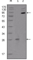
Figure 1: Western blot analysis using EphB2 mouse mAb against truncated EphB2 recombinant protein (1) and extracellular EphB2(aa19-476)-hIgGFc transfected CHO-K1 cell lysate(2).
Immunofluorescence analysis

Figure 2:Immunofluorescence analysis of Hela (left) and HepG2 (right) cells using EphB2 mouse mAb (green). Red: Actin filaments have been labeled with DY-554 phalloidin. Blue: DRAQ5 fluorescent DNA dye.
For Research Use Only. Not for use in diagnostic procedures.