
EPCAM Primary Antibody
Item Information
Catalog #
Size
Price
Description
This gene encodes a carcinoma-associated antigen and is a member of a family that includes at least two type I membrane proteins. This antigen is expressed on most normal epithelial cells and gastrointestinal carcinomas and functions as a homotypic calcium-independent cell adhesion molecule. The antigen is being used as a target for immunotherapy treatment of human carcinomas. Mutations in this gene result in congenital tufting enteropathy. [provided by RefSeq, Dec 2008]
Product Overview
Entrez GenelD
4072
Aliases
ESA; KSA; M4S1; MK-1; DIAR5; EGP-2; EGP40; KS1/4; MIC18; TROP1; EGP314; HNPCC8; TACSTD1
Clone#
4D6E7
Host / Isotype
Mouse / Mouse IgG2a
Immunogen
Purified recombinant fragment of human EPCAM (AA: extra(116-265)) expressed in E. Coli.
Formulation
Purified antibody in PBS with 0.05% sodium azide
Storage
Store at 4°C short term. Aliquot and store at -20°C long term. Avoid freeze/thaw cycles.
Product Applications
WB (Western Blot)
1/500 - 1/2000
IHC_P(Immunohistochemistry)
1/200-1/1000
FCM (Flow Cytometry)
1/200-1/400
ELISA
1/10000
References
1,Asian Pac J Cancer Prev. 2020 Mar 1;21(3):861-866. 2,J Radiat Res. 2019 Nov 22;60(6):803-811.
Product Image
ELISA
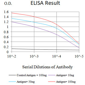
Figure 1: Black line: Control Antigen (100 ng);Purple line: Antigen (10ng); Blue line: Antigen (50 ng); Red line: Antigen (100 ng)
WESTERN BLOT
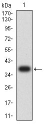
Figure 2: Western blot analysis using EPCAM mAb against human EPCAM (AA: extra(116-265)) recombinant protein. (Expected MW is 38 kDa)
WESTERN BLOT
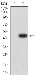
Figure 3: Western blot analysis using EPCAM mAb against HEK293-6e (1) and EPCAM (AA: extra(116-265))-hIgGFc transfected HEK293-6e (2) cell lysate.
WESTERN BLOT
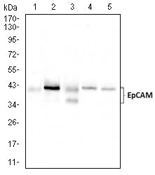
Figure 4: Western blot analysis using EPCAM mouse mAb against HCT116 (1), HT-29 (2),SW480 (3),Sw-620 (4) , and T47D (5) cell lysate.
FLOW CYTOMETRY
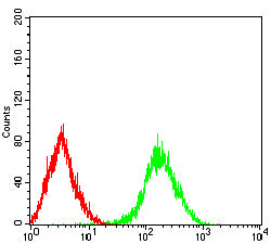
Figure 5: Flow cytometric analysis of Lovo cells using EPCAM mouse mAb (green) and negative control (red).
IMMUNOHISTOCHEMISTRY

Figure 6: Immunohistochemical analysis of paraffin-embedded cervical cancer tissues using EPCAM mouse mAb with DAB staining.
IMMUNOHISTOCHEMISTRY
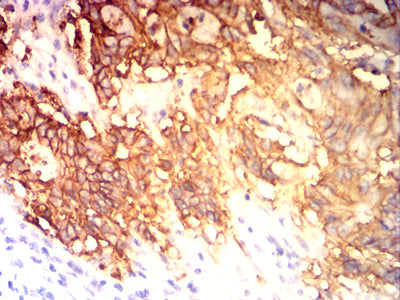
Figure 7: Immunohistochemical analysis of paraffin-embedded colon cancer tissues using EPCAM mouse mAb with DAB staining.
For Research Use Only. Not for use in diagnostic procedures.