

EPCAM Primary Antibody
Item Information
Catalog #
Size
Price
Description
This gene encodes a carcinoma-associated antigen and is a member of a family that includes at least two type I membrane proteins. This antigen is expressed on most normal epithelial cells and gastrointestinal carcinomas and functions as a homotypic calcium-independent cell adhesion molecule. The antigen is being used as a target for immunotherapy treatment of human carcinomas. Mutations in this gene result in congenital tufting enteropathy.
Product Overview
Entrez GenelD
4072
Aliases
HOX2; HOX2F; HOX-2.6
Clone#
3H6A6
Host / Isotype
Mouse / IgG1
Species Reactivity
Human
Immunogen
Purified recombinant fragment of human EPCAM (AA: Extra(24-265)) expressed in E. Coli.
Formulation
Purified antibody in PBS with 0.05% sodium azide.
Storage
Store at 4°C short term. Aliquot and store at -20°C long term. Avoid freeze/thaw cycles.
Product Applications
WB (Western Blot)
1/500 - 1/2000
FCM (Flow Cytometry)
1/200 - 1/400
ELISA
1/10000
References
1. BMC Cancer. 2012 Oct 30;12:501.
2. Am J Surg Pathol. 2012 Dec;36(12):1809-16.
2. Am J Surg Pathol. 2012 Dec;36(12):1809-16.
Product Image
Western Blot

Figure 1: Western blot analysis using EPCAM mAb against human EPCAM (AA: Extra(24-265)) recombinant protein. (Expected MW is 53.4 kDa)
Western Blot

Figure 2: Western blot analysis using EPCAM mAb against HEK293 (1) and EPCAM (AA: Extra(24-265))-hIgGFc transfected HEK293 (2) cell lysate.
Western Blot

Figure 3: Western blot analysis using EPCAM mouse mAb against A431 (1), MCF-7 (2) cell lysate.
Flow cytometric

Figure 5: Flow cytometric analysis of A431 cells using EPCAM mouse mAb (green) and negative control (red).
Elisa
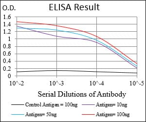
Black line: Control Antigen (100 ng); Purple line: Antigen(10ng); Blue line: Antigen (50 ng); Red line: Antigen (100 ng);
For Research Use Only. Not for use in diagnostic procedures.