
EMD Primary Antibody
Item Information
Catalog #
Size
Price
Description
Emerin is a serine-rich nuclear membrane protein and a member of the nuclear lamina-associated protein family. It mediates membrane anchorage to the cytoskeleton. Dreifuss-Emery muscular dystrophy is an X-linked inherited degenerative myopathy resulting from mutation in the emerin gene.
Product Overview
Entrez GenelD
2010
Aliases
STA; EDMD; LEMD5
Clone#
8F5A8
Host / Isotype
Mouse / IgG1
Species Reactivity
Human
Immunogen
Purified recombinant fragment of human EMD (AA: 1-222) expressed in E. Coli.
Formulation
Purified antibody from tissue culture in PBS with 0.05% sodium azide
Storage
Store at 4°C short term. Aliquot and store at -20°C long term. Avoid freeze/thaw cycles.
Product Applications
WB (Western Blot)
1/500 - 1/2000
IHC_P(Immunohistochemistry)
1/200 - 1/1000
ICC (Immunocytochemistry)
1/200 - 1/1000
FCM (Flow Cytometry)
1/200 - 1/400
ELISA
1/10000
References
1.Histopathology. 2009 Apr;54(5):571-9.
2.J Cell Biol. 2007 Sep 10;178(6):897-904.
2.J Cell Biol. 2007 Sep 10;178(6):897-904.
Product Image
Elisa
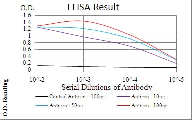
Figure 1: Black line: Control Antigen (100 ng); Purple line: Antigen(10ng); Blue line: Antigen (50 ng); Red line: Antigen (100 ng);
Western Blot
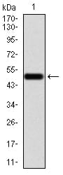
Figure 2:Western blot analysis using EMD mAb against human EMD (AA: 1-222) recombinant protein. (Expected MW is 51.1 kDa)
Western Blot
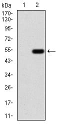
Figure 3:Western blot analysis using EMD mAb against HEK293 (1) and EMD (AA: 1-222)-hIgGFc transfected HEK293 (2) cell lysate.
Immunofluorescence analysis
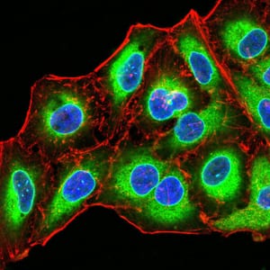
Figure 4:Immunofluorescence analysis of Hela cells using EMD mouse mAb (green). Blue: DRAQ5 fluorescent DNA dye. Red: Actin filaments have been labeled with Alexa Fluor- 555 phalloidin. Secondary antibody from Fisher (Cat#: 35503)
Flow cytometric

Figure 5:Flow cytometric analysis of A549 cells using EMD mouse mAb (green) and negative control (red).
Flow cytometric

Figure 6:Flow cytometric analysis of K562 cells using EMD mouse mAb (green) and negative control (red).
Immunohistochemical analysis
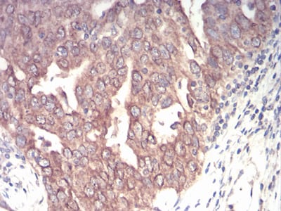
Figure 7:Immunohistochemical analysis of paraffin-embedded ovarian cancer tissues using EMD mouse mAb with DAB staining.
For Research Use Only. Not for use in diagnostic procedures.