
ELK1 Primary Antibody
Item Information
Catalog #
Size
Price
Description
The transcription factor ELK1 is a family of member of ETS oncogene family and of the ternary complex factor (TCF) subfamily,which is located on chromosome Xp11.2 and stimulates
transcription. binds to purine-rich DNA sequences. Proteins of the TCF subfamily form a ternary complex by binding to the the serum response factor and the serum reponse element in the promoter of the c-fos proto-oncogene. The protein encoded by this gene is a nuclear target for the ras-raf-MAPK signaling cascade.
Elk1 is phosphorylated by MAP kinase pathways at a cluster of S/T motifs at its C terminus,It appears to be a direct target of activated MAP kinase. Biochemical studies indicate that Elk1 is a good substrate for MAP kinase, the kinetics of Elk1phosphorylation and activation correlate with MAP kinase activity, and interfering mutants of MAP kinase block Elk1 activation in vivo. More recent studies have shown that Elk1 is also a target of the Stress Activated Kinase SAPK/JNK. Phosphorylation of Elk1 has also been implicated in synaptic plasticity in the adult hippocampus.
Product Overview
Entrez GenelD
2002
Aliases
ELK1
Clone#
3H6D12
Host / Isotype
Mouse / IgG1
Species Reactivity
Human
Immunogen
Purified recombinant fragment of ELK1 expressed in E. Coli.
Formulation
Ascitic fluid containing 0.03% sodium azide.
Storage
Store at 4°C short term. Aliquot and store at -20°C long term. Avoid freeze/thaw cycles.
Product Applications
WB (Western Blot)
1/500 - 1/2000
IHC_P(Immunohistochemistry)
1/200 - 1/1000
ELISA
1/10000
References
1. Rao,V.N., et al. 1989.Science.244 (4900):66-70.
2. Hsieh,Y.H., et al. 2006.Biochem. Biophys. Res. Commun. 339 (1): 217-225.
3. Gille,H., Strahl,T. and Shaw,P.E.1995. Curr. Biol. 5 (10): 1191-1200.
4. Gille,H., et al. 1995.EMBO J. 14 (5): 951-962.
2. Hsieh,Y.H., et al. 2006.Biochem. Biophys. Res. Commun. 339 (1): 217-225.
3. Gille,H., Strahl,T. and Shaw,P.E.1995. Curr. Biol. 5 (10): 1191-1200.
4. Gille,H., et al. 1995.EMBO J. 14 (5): 951-962.
Product Image
Western Blot
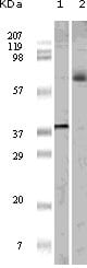
Figure 1: Western blot analysis using ELK1 mouse mAb against truncated ELK1 recombinant protein (1) and K562 cell lysate (2).
Immunohistochemical analysis

Figure 2: Immunohistochemical analysis of paraffin-embedded human brain tumor tissue, showing nuclear and cytoplasmic localization using ELK1 mouse mAb with DAB staining.
For Research Use Only. Not for use in diagnostic procedures.