

ELANE Primary Antibody
Item Information
Catalog #
Size
Price
Description
Elastases form a subfamily of serine proteases that hydrolyze many proteins in addition to elastin. Humans have six elastase genes which encode structurally similar proteins. The encoded preproprotein is proteolytically processed to generate the active protease. Following activation, this protease hydrolyzes proteins within specialized neutrophil lysosomes, called azurophil granules, as well as proteins of the extracellular matrix. The enzyme may play a role in degenerative and inflammatory diseases through proteolysis of collagen-IV and elastin. This protein also degrades the outer membrane protein A (OmpA) of E. coli as well as the virulence factors of such bacteria as Shigella, Salmonella and Yersinia. Mutations in this gene are associated with cyclic neutropenia and severe congenital neutropenia (SCN). This gene is present in a gene cluster on chromosome 19.
Product Overview
Entrez GenelD
1991
Aliases
GE; NE; HLE; HNE; ELA2; SCN1; PMN-E
Clone#
6B6B10
Host / Isotype
Mouse / IgG1
Species Reactivity
Human, Monkey
Immunogen
Purified recombinant fragment of human ELANE (AA: 140-267) expressed in E. Coli.
Formulation
Purified antibody in PBS with 0.05% sodium azide
Storage
Store at 4°C short term. Aliquot and store at -20°C long term. Avoid freeze/thaw cycles.
Product Applications
WB (Western Blot)
1/500 - 1/2000
IHC_P(Immunohistochemistry)
1/200 - 1/1000
FCM (Flow Cytometry)
1/200 - 1/400
ELISA
1/10000
References
1.Pediatr Blood Cancer. 2015 Dec;62(12):2229-31.
2.Hum Mutat. 2013 Jun;34(6):905-14.
2.Hum Mutat. 2013 Jun;34(6):905-14.
Product Image
Elisa
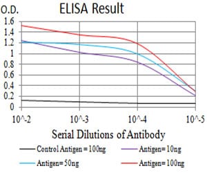
Figure 1: Black line: Control Antigen (100 ng);Purple line: Antigen (10ng); Blue line: Antigen (50 ng); Red line:Antigen (100 ng)
Western Blot
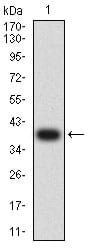
Figure 2:Western blot analysis using ELANE mAb against human ELANE (AA: 140-267) recombinant protein. (Expected MW is 39 kDa)
Western Blot
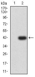
Figure 3:Western blot analysis using ELANE mAb against HEK293 (1) and ELANE (AA: 140-267)-hIgGFc transfected HEK293 (2) cell lysate.
Western Blot
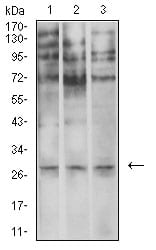
Figure 4:Western blot analysis using ELANE mouse mAb against U937 (1), SPC-A-1 (2), and COS7 (3) cell lysate.
Flow cytometric
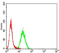
Figure 5:Flow cytometric analysis of Hela cells using ELANE mouse mAb (green) and negative control (red).
Immunohistochemical analysis
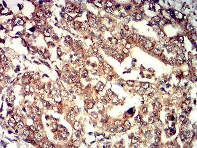
Figure 6:Immunohistochemical analysis of paraffin-embedded stomach cancer tissues using ELANE mouse mAb with DAB staining.
For Research Use Only. Not for use in diagnostic procedures.