

EIF5A Primary Antibody
Item Information
Catalog #
Size
Price
Description
EIF5A (eukaryotic translation initiation factor 5A) is a protein-coding gene. Diseases associated with EIF5A include lung adenocarcinoma, and intrahepatic cholangiocarcinoma, and among its related super-pathways are Post-translational protein modification and Apoptotic Pathways in Synovial Fibroblasts. GO annotations related to this gene include ribosome binding and RNA binding. An important paralog of this gene is EIF5AL1.
Product Overview
Entrez GenelD
1984
Aliases
EIF-5A; EIF5A1; eIF5AI
Clone#
4E10G8
Host / Isotype
Mouse / IgG1
Species Reactivity
Human
Immunogen
Purified recombinant fragment of human EIF5A (AA: full(1-154)) expressed in E. Coli.
Formulation
Purified antibody from tissue culture in PBS with 0.05% sodium azide
Storage
Store at 4°C short term. Aliquot and store at -20°C long term. Avoid freeze/thaw cycles.
Product Applications
WB (Western Blot)
1/500 - 1/2000
IHC_P(Immunohistochemistry)
1/200 - 1/1000
ICC (Immunocytochemistry)
1/200 - 1/1000
FCM (Flow Cytometry)
1/200 - 1/400
ELISA
1/10000
References
1. Int J Cancer. 2011 Jul 1;129(1):143-50.
2. Int J Cancer. 2010 Aug 15;127(4):968-76.
2. Int J Cancer. 2010 Aug 15;127(4):968-76.
Product Image
Western Blot
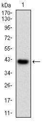
Figure 1: Western blot analysis using EIF5A mAb against human EIF5A (AA: full(1-154)) recombinant protein. (Expected MW is 42.8 kDa)
Western Blot
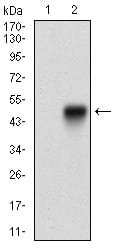
Figure 2: Western blot analysis using EIF5A mAb against HEK293 (1) and EIF5A (AA: full(1-154))-hIgGFc transfected HEK293 (2) cell lysate.
Immunofluorescence analysis
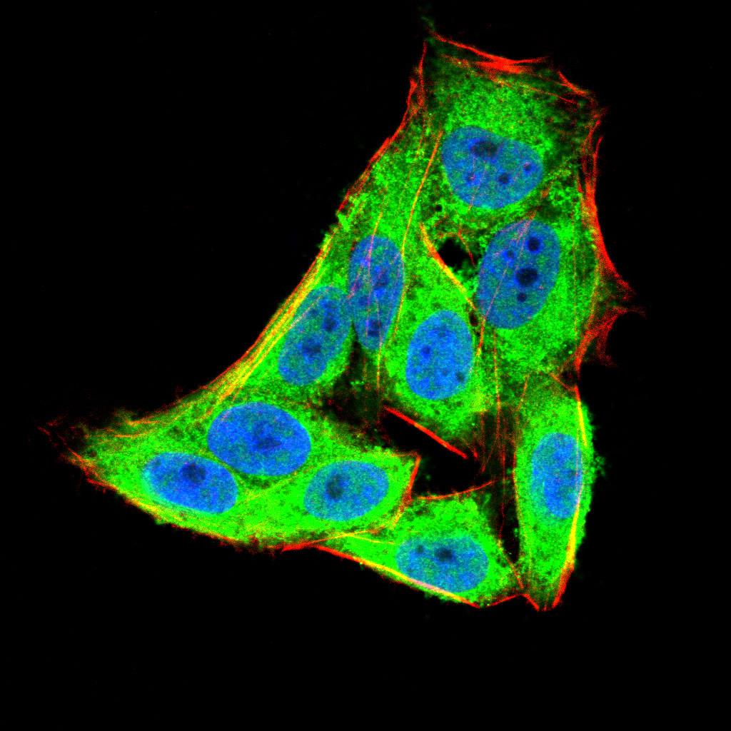
Figure 3:Immunofluorescence analysis of Hela cells using EIF5A mouse mAb (green). Blue: DRAQ5 fluorescent DNA dye. Red: Actin filaments have been labeled with Alexa Fluor- 555 phalloidin. Secondary antibody from Fisher (Cat#: 35503)
Flow cytometric
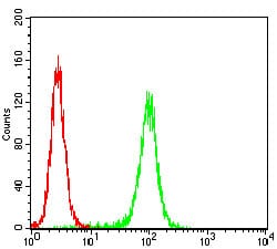
Figure 4: Flow cytometric analysis of Hela cells using EIF5A mouse mAb (green) and negative control (red).
Immunohistochemical analysis
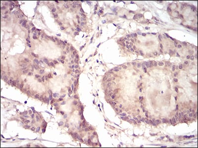
Figure 5: Immunohistochemical analysis of paraffin-embedded colon cancer tissues using EIF5A mouse mAb with DAB staining.
Immunohistochemical analysis
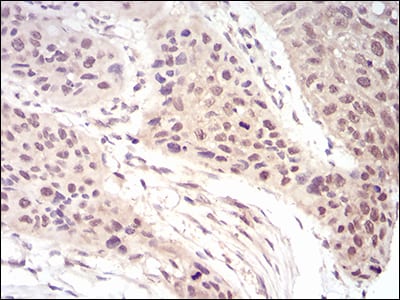
Figure 6: Immunohistochemical analysis of paraffin-embedded esophageal cancer tissues using EIF5A mouse mAb with DAB staining.
Elisa
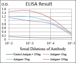
Black line: Control Antigen (100 ng); Purple line: Antigen(10ng); Blue line: Antigen (50 ng); Red line: Antigen (100 ng);
For Research Use Only. Not for use in diagnostic procedures.