

EIF5 Primary Antibody
Item Information
Catalog #
Size
Price
Description
Eukaryotic translation initiation factor-5 (EIF5) interacts with the 40S initiation complex to promote hydrolysis of bound GTP with concomitant joining of the 60S ribosomal subunit to the 40S initiation complex. The resulting functional 80S ribosomal initiation complex is then active in peptidyl transfer and chain elongations (summary by Si et al., 1996 [PubMed 8663286]).
Product Overview
Entrez GenelD
1983
Aliases
EIF-5; EIF-5A
Clone#
6D6H11
Host / Isotype
Mouse / IgG2a
Species Reactivity
Human
Immunogen
Purified recombinant fragment of human EIF5 (AA: 1-300) expressed in E. Coli.
Formulation
Purified antibody in PBS with 0.05% sodium azide
Storage
Store at 4°C short term. Aliquot and store at -20°C long term. Avoid freeze/thaw cycles.
Product Applications
WB (Western Blot)
1/500 - 1/2000
IHC_P(Immunohistochemistry)
1/200 - 1/1000
ICC (Immunocytochemistry)
1/50 - 1/250
FCM (Flow Cytometry)
1/200 - 1/400
ELISA
1/10000
References
1.Biochemistry. 2006 Apr 11;45(14):4550-8.
2.Biochem Biophys Res Commun. 2011 Dec 2;415(4):567-72.
2.Biochem Biophys Res Commun. 2011 Dec 2;415(4):567-72.
Product Image
Elisa

Figure 1: Black line: Control Antigen (100 ng);Purple line: Antigen (10ng); Blue line: Antigen (50 ng); Red line:Antigen (100 ng)
Western Blot

Figure 2:Western blot analysis using EIF5 mAb against human EIF5 (AA: 1-300) recombinant protein. (Expected MW is 60.2 kDa)
Western Blot

Figure 3:Western blot analysis using EIF5 mAb against HEK293 (1) and EIF5 (AA: 1-300)-hIgGFc transfected HEK293 (2) cell lysate.
Immunofluorescence analysis

Figure 4:Immunofluorescence analysis of Hela cells using EIF5 mouse mAb (green). Blue: DRAQ5 fluorescent DNA dye. Red: Actin filaments have been labeled with Alexa Fluor- 555 phalloidin. Secondary antibody from Fisher (Cat#: 35503)
Flow cytometric

Figure 5:Flow cytometric analysis of Hela cells using EIF5 mouse mAb (green) and negative control (red).
Immunohistochemical analysis

Figure 6:Immunohistochemical analysis of paraffin-embedded bladder cancer tissues using EIF5 mouse mAb with DAB staining.
Immunohistochemical analysis
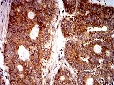
Figure 7:Immunohistochemical analysis of paraffin-embedded rectum cancer tissues using EIF5 mouse mAb with DAB staining.
For Research Use Only. Not for use in diagnostic procedures.