

EIF4B Primary Antibody
Item Information
Catalog #
Size
Price
Description
The eukaryotic translation initiation factor 4B (eIF4B) plays a critical role in recruiting the 40S ribosomal subunit to the mRNA.It functions in close association with eIF4F and eIF4A. It binds near the 5'-terminal cap of mRNA in the presence of eIF4F and ATP. It promotes the ATPase activity and the ATP-dependent RNA unwinding activity of both eIF4A and eIF4F
Product Overview
Entrez GenelD
1975
Aliases
EIF-4B; PRO1843
Clone#
1F5
Host / Isotype
Mouse / IgG2b
Species Reactivity
Human, Mouse
Immunogen
Purified recombinant fragment of human EIF4B expressed in E. Coli.
Formulation
Purified antibody in PBS with 0.05% sodium azide
Storage
Store at 4°C short term. Aliquot and store at -20°C long term. Avoid freeze/thaw cycles.
Product Applications
WB (Western Blot)
1/500 - 1/2000
IHC_P(Immunohistochemistry)
1/200 - 1/1000
ICC (Immunocytochemistry)
1/200 - 1/1000
FCM (Flow Cytometry)
1/200 - 1/400
ELISA
1/10000
References
1. Mol Biol Cell. 2009 Jun;20(11):2673-83.
2. EMBO J. 2006 Jun 21;25(12):2781-91.
2. EMBO J. 2006 Jun 21;25(12):2781-91.
Product Image
Western Blot

Figure 1: Western blot analysis using EIF4B mAb against human EIF4B (AA: 381-585) recombinant protein. (Expected MW is 48.3 kDa)
Western Blot

Figure 2: Western blot analysis using EIF4B mouse mAb against A549 (1), A431 (2), HepG2 (3), HEK293 (4), HeLa (5), Jurkat (6), K562 (7), NIH3T3 (8), and MCF-7 (9) cell lysate.
Flow cytometric

Figure 4: Flow cytometric analysis of HeLa cells using EIF4B mouse mAb (green) and negative control (red).
Immunofluorescence analysis
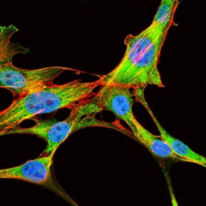
Figure 5 : Immunofluorescence analysis of NIH3T3 cells using EIF4B mouse mAb (green). Blue: DRAQ5 fluorescent DNA dye. Red: Actin filaments have been labeled with Alexa Fluor-555 phalloidin.
Immunohistochemical analysis

Figure 5: Immunohistochemical analysis of paraffin-embedded colon cancer tissues using EIF4B mouse mAb with DAB staining.
Elisa

Black line: Control Antigen (100 ng); Purple line: Antigen(10ng); Blue line: Antigen (50 ng); Red line: Antigen (100 ng);
For Research Use Only. Not for use in diagnostic procedures.