EhpB6 Primary Antibody

Item Information
Catalog #
Size
Price
Description

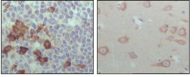
EhpB6: EPH receptor B6. Ephrin receptors and their ligands, the ephrins, mediate numerous developmental processes, particularly in the nervous system. Based on their structures and sequence relationships, ephrins are divided into the ephrin-A (EFNA) class, which are anchored to the membrane by a glycosylphosphatidylinositol linkage, and the ephrin-B (EFNB) class, which are transmembrane proteins. The Eph family of receptors are divided into 2 groups based on the similarity of their extracellular domain sequences and their affinities for binding ephrin-A and ephrin-B ligands. Ephrin receptors make up the largest subgroup of the receptor tyrosine kinase (RTK) family. The ephrin receptor encoded by this gene lacks the kinase activity of most receptor tyrosine kinases and binds to ephrin-B ligands.

Product Overview
Entrez GenelD
2051
Aliases
HEP; EPHB6
Clone#
2A6B9
Host / Isotype
Mouse / IgG1
Species Reactivity
Human
Immunogen
Purified recombinant fragment of EphB6 (aa601-750) expressed in E. Coli.
Formulation
Ascitic fluid containing 0.03% sodium azide.
Storage
4°C; -20°C for long term storage
Product Applications
WB (Western Blot)
1/500 - 1/2000
IHC_P(Immunohistochemistry)
1/200 - 1/1000
ELISA
1/10000
References
1. J Clin Invest. 2002 Oct;110(8):1141-50.
2. J Biol Chem. 2002 Feb 8;277(6):3823-8. Epub 2001 Nov 16.
3. J Biol Chem. 2003 Mar 21;278(12):10150-6. Epub 2003 Jan 6.
4. Biochem Biophys Res Commun. 2006 Feb 3;340(1):268-76.
Product Image
Western Blot
Figure 1: Western blot analysis using EhpB6 mouse mAb against truncated EhpB6 recombinant protein (1).
Immunohistochemical analysis
Figure 2: Immunohistochemical analysis of paraffin-embedded human lymph node (left) and brain (right), showing cytoplasmic localization with DAB staining using EhpB6 mouse mAb.
For Research Use Only. Not for use in diagnostic procedures.