
EHMT2 Primary Antibody
Item Information
Catalog #
Size
Price
Description
This gene encodes a methyltransferase that methylates lysine residues of histone H3. Methylation of H3 at lysine 9 by this protein results in recruitment of additional epigenetic regulators and repression of transcription. This gene was initially thought to be two different genes, NG36 and G9a, adjacent to each other in the HLA locus. Alternative splicing results in multiple transcript variants.
Product Overview
Entrez GenelD
10919
Aliases
G9A; BAT8; GAT8; NG36; KMT1C; C6orf30
Clone#
2D12A11
Host / Isotype
Mouse / IgG1
Species Reactivity
Human
Immunogen
Purified recombinant fragment of human EHMT2 (AA: 317-471) expressed in E. Coli.
Formulation
Purified antibody in PBS with 0.05% sodium azide
Storage
Store at 4°C short term. Aliquot and store at -20°C long term. Avoid freeze/thaw cycles.
Product Applications
WB (Western Blot)
1/500 - 1/2000
IHC_P(Immunohistochemistry)
1/200 - 1/1000
ICC (Immunocytochemistry)
1/200 - 1/1000
FCM (Flow Cytometry)
1/200 - 1/400
ELISA
1/10000
References
1.Proc Natl Acad Sci U S A. 2017 Jul 3;114(27):7077-7082.
2.Mol Med Rep. 2016 Nov;14(5):4613-4621.
2.Mol Med Rep. 2016 Nov;14(5):4613-4621.
Product Image
Elisa
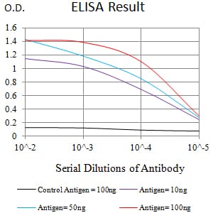
Figure 1:Black line: Control Antigen (100 ng);Purple line: Antigen (10ng); Blue line: Antigen (50 ng); Red line:Antigen (100 ng)
Western Blot
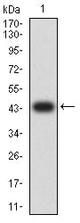
Figure 2:Western blot analysis using EHMT2 mAb against human EHMT2 (AA: 317-471) recombinant protein. (Expected MW is 44 kDa)
Western Blot
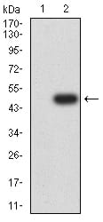
Figure 3:Western blot analysis using EHMT2 mAb against HEK293 (1) and EHMT2 (AA: 317-471)-hIgGFc transfected HEK293 (2) cell lysate.
Immunofluorescence analysis
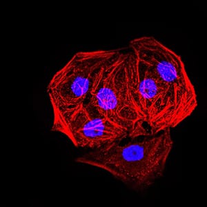
Figure 4:Immunofluorescence analysis of Hela cells. Blue: DRAQ5 fluorescent DNA dye. Red: Actin filaments have been labeled with Alexa Fluor- 555 phalloidin.
Immunofluorescence analysis
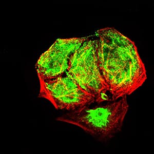
Figure 5:Immunofluorescence analysis of Hela cells using EHMT2 mouse mAb (green). Blue: DRAQ5 fluorescent DNA dye. Red: Actin filaments have been labeled with Alexa Fluor- 555 phalloidin. Secondary antibody from Fisher (Cat#: 35503)
Flow cytometric
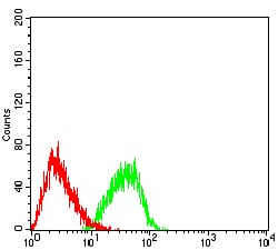
Figure 6:Flow cytometric analysis of HL-60 cells using EHMT2 mouse mAb (green) and negative control (red).
Immunohistochemical analysis
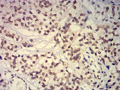
Figure 7:Immunohistochemical analysis of paraffin-embedded bladder cancer tissues using EHMT2 mouse mAb with DAB staining.
Immunohistochemical analysis
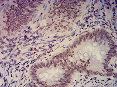
Figure 8:Immunohistochemical analysis of paraffin-embedded rectum cancer tissues using EHMT2 mouse mAb with DAB staining.
For Research Use Only. Not for use in diagnostic procedures.