

DOC2 Primary Antibody
Item Information
Catalog #
Size
Price
Description
This gene encodes a mitogen-responsive phosphoprotein. It is expressed in normal ovarian epithelial cells, but is down-regulated or absent from ovarian carcinoma cell lines, suggesting its role as a tumor suppressor. This protein binds to the SH3 domains of GRB2, an adaptor protein that couples tyrosine kinase receptors to SOS (a guanine nucleotide exchange factor for Ras), via its C-terminal proline-rich sequences, and may thus modulate growth factor/Ras pathways by competing with SOS for binding to GRB2. Alternatively spliced transcript variants encoding different isoforms have been found for this gene.
Product Overview
Entrez GenelD
1601
Aliases
DAB2; DOC-2
Clone#
2H7C4
Host / Isotype
Mouse / IgG1
Species Reactivity
Human
Immunogen
Purified recombinant fragment of human DOC2 (AA: 652-749) expressed in E. Coli.
Formulation
Purified antibody in PBS with 0.05% sodium azide
Storage
Store at 4°C short term. Aliquot and store at -20°C long term. Avoid freeze/thaw cycles.
Product Applications
WB (Western Blot)
1/500 - 1/2000
ICC (Immunocytochemistry)
1/200 - 1/1000
FCM (Flow Cytometry)
1/200 - 1/400
ELISA
1/10000
References
1.Mol Biol Cell. 2014 May;25(10):1620-8.
2.Exp Cell Res. 2012 Mar 10;318(5):550-7.
2.Exp Cell Res. 2012 Mar 10;318(5):550-7.
Product Image
Elisa
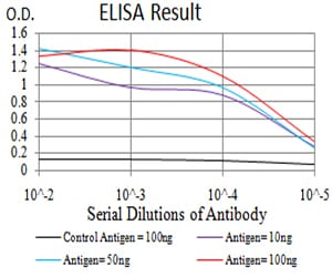
Figure 1: Black line: Control Antigen (100 ng);Purple line: Antigen (10ng); Blue line: Antigen (50 ng); Red line:Antigen (100 ng)
Western Blot
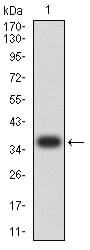
Figure 2:Western blot analysis using DOC2 mAb against human DOC2 (AA: 652-749) recombinant protein. (Expected MW is 36.6 kDa)
Western Blot
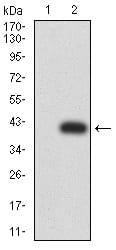
Figure 3:Western blot analysis using DOC2 mAb against HEK293 (1) and DOC2 (AA: 652-749)-hIgGFc transfected HEK293 (2) cell lysate.
Immunofluorescence analysis
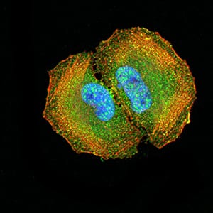
Figure 4:Immunofluorescence analysis of Hela cells using DOC2 mouse mAb (green). Blue: DRAQ5 fluorescent DNA dye. Red: Actin filaments have been labeled with Alexa Fluor- 555 phalloidin. Secondary antibody from Fisher (Cat#: 35503)
Flow cytometric
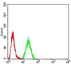
Figure 5:Flow cytometric analysis of Hela cells using DOC2 mouse mAb (green) and negative control (red).
For Research Use Only. Not for use in diagnostic procedures.