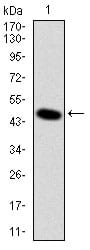
DLK1 Primary Antibody

Item Information
Catalog #
Size
Price
Description

This gene encodes a transmembrane protein containing six epidermal growth factor repeats. The protein is involved in the differentiation of several cell types, including adipocytes; it is also thought to be a tumor suppressor. It is one of several imprinted genes located in a region of on chr 14q32. Certain mutations in this imprinted region can cause phenotypes similar to maternal and paternal uniparental disomy of chromosome 14 (UPD14). This gene is expressed from the paternal allele. A polymorphism within this gene has been associated with child and adolescent obesity. The mode of inheritance for this polymorphism is polar overdominance; this non-Mendelian inheritance pattern was first described in sheep with the callipyge phenotype, which is characterized by muscle hypertrophy and decreased fat mass.

Product Overview
Entrez GenelD
8788
Aliases
DLK; FA1; ZOG; pG2; PREF1; Pref-1
Clone#
3A10
Host / Isotype
Mouse / IgG1
Species Reactivity
Human
Immunogen
Purified recombinant fragment of human DLK1 expressed in E. Coli.
Formulation
Ascitic fluid containing 0.03% sodium azide.
Storage
4°C; -20°C for long term storage
Product Applications
WB (Western Blot)
1/500 - 1/2000
ICC (Immunocytochemistry)
1/200 - 1/1000
FCM (Flow Cytometry)
1/200 - 1/400
ELISA
1/10000
References
1. Epigenetics. 2009 Oct 1;4(7):469-75.
2. Mol Biol Cell. 2009 Jul;20(14):3353-62. 
Product Image
Western Blot
Figure 1: Western blot analysis using DLK1 mAb against human DLK1 (AA: 174-349) recombinant protein. (Expected MW is 44.9 kDa)
Immunofluorescence analysis
Figure 2: Immunofluorescence analysis of U251 cells using DLK1 mouse mAb (green). Blue: DRAQ5 fluorescent DNA dye. Red: Actin filaments have been labeled with Alexa Fluor-555 phalloidin.
Flow cytometric
Figure 3: Flow cytometric analysis of NIH/3T3 cells using DLK1 mouse mAb (green) and negative control (red).
Elisa
Black line: Control Antigen (100 ng); Purple line: Antigen(10ng); Blue line: Antigen (50 ng); Red line: Antigen (100 ng);
For Research Use Only. Not for use in diagnostic procedures.