

DLG4 Primary Antibody
Item Information
Catalog #
Size
Price
Description
This gene encodes a member of the membrane-associated guanylate kinase (MAGUK) family. It heteromultimerizes with another MAGUK protein, DLG2, and is recruited into NMDA receptor and potassium channel clusters. These two MAGUK proteins may interact at postsynaptic sites to form a multimeric scaffold for the clustering of receptors, ion channels, and associated signaling proteins. Multiple transcript variants encoding different isoforms have been found for this gene.
Product Overview
Entrez GenelD
1742
Aliases
MRD62; PSD95; SAP90; SAP-90
Clone#
2D5H2
Host / Isotype
Mouse / Mouse IgG1
Species Reactivity
Human, Mouse, Rat
Immunogen
Purified recombinant fragment of human DLG4 (AA: 54-300) expressed in HEK293-6e cells supernatant.
Formulation
Purified antibody in PBS with 0.05% sodium azide
Storage
Store at 4°C short term. Aliquot and store at -20°C long term. Avoid freeze/thaw cycles.
Product Applications
WB (Western Blot)
1/500 - 1/2000
IHC_P(Immunohistochemistry)
1/200 - 1/1000
FCM (Flow Cytometry)
1/200 - 1/400
ELISA
1/10000
References
1.J Mol Neurosci. 2020 Sep;70(9):1389-1402.
2.J Biol Chem. 2020 Feb 14;295(7):1992-2000.
2.J Biol Chem. 2020 Feb 14;295(7):1992-2000.
Product Image
Elisa
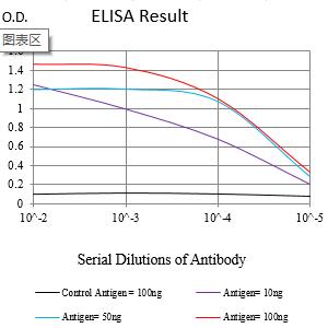
Figure 1:Black line: Control Antigen (100 ng);Purple line: Antigen (10ng); Blue line: Antigen (50 ng); Red line:Antigen (100 ng)
Western Blot

Figure 2:Western blot analysis using DLG4 mAb against human DLG4 (AA: 54-300) recombinant protein. (Expected MW is 57 kDa)
Western Blot

Figure 4:Western blot analysis using DLG4 mouse mAb against Mouse brain (1), and Rat brain (2) tissue lysate.
Immunofluorescence analysis

Figure 5:Flow cytometric analysis of LNCAP cells using DLG4 mouse mAb (green) and negative control (red).
Immunohistochemical analysis
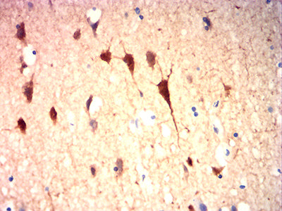
Figure 6:Immunohistochemical analysis of paraffin-embedded human brain tissues using DLG4 mouse mAb with DAB staining.
For Research Use Only. Not for use in diagnostic procedures.