

DDX5 Primary Antibody
Item Information
Catalog #
Size
Price
Description
DEAD box proteins, characterized by the conserved motif Asp-Glu-Ala-Asp (DEAD), are putative RNA helicases. They are implicated in a number of cellular processes involving alteration of RNA secondary structure, such as translation initiation, nuclear and mitochondrial splicing, and ribosome and spliceosome assembly. Based on their distribution patterns, some members of this family are believed to be involved in embryogenesis, spermatogenesis, and cellular growth and division. This gene encodes a DEAD box protein, which is a RNA-dependent ATPase, and also a proliferation-associated nuclear antigen, specifically reacting with the simian virus 40 tumor antigen. This gene consists of 13 exons, and alternatively spliced transcripts containing several intron sequences have been detected, but no isoforms encoded by these transcripts have been identified.
Product Overview
Entrez GenelD
1655
Aliases
p68; HLR1; G17P1; HUMP68
Clone#
4F1C12
Host / Isotype
Mouse / IgG2a
Species Reactivity
Human, Mouse, Monkey
Immunogen
Purified recombinant fragment of human DDX5 (AA: 475-614) expressed in E. Coli.
Formulation
Purified antibody in PBS with 0.05% sodium azide
Storage
Store at 4°C short term. Aliquot and store at -20°C long term. Avoid freeze/thaw cycles.
Product Applications
WB (Western Blot)
1/500 - 1/2000
IHC_P(Immunohistochemistry)
1/200 - 1/1000
FCM (Flow Cytometry)
1/200 - 1/400
ELISA
1/10000
References
1.Cancer Discov. 2012 Sep;2(9):812-25.
2.Nucleic Acids Res. 2012 Apr;40(7):3159-71.
2.Nucleic Acids Res. 2012 Apr;40(7):3159-71.
Product Image
Elisa
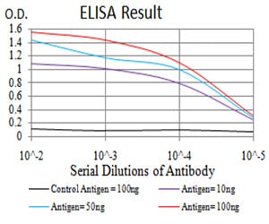
Figure 1: Black line: Control Antigen (100 ng);Purple line: Antigen (10ng); Blue line: Antigen (50 ng); Red line:Antigen (100 ng)
Western Blot

Figure 2:Western blot analysis using DDX5 mAb against human DDX5 (AA: 475-614) recombinant protein. (Expected MW is 41.5 kDa)
Western Blot

Figure 3:Western blot analysis using DDX5 mAb against HEK293 (1) and DDX5 (AA: 475-614)-hIgGFc transfected HEK293 (2) cell lysate.
Western Blot

Figure 4:Western blot analysis using DDX5 mouse mAb against HT-29 (1), Hela (2), NIH/3T3 (3), COS7 (4), SW620 (5), Jurkat (6), A431 (7), and MCF-7 (8) cell lysate.
Flow cytometric

Figure 5:Flow cytometric analysis of Hela cells using DDX5 mouse mAb (green) and negative control (red).
Immunohistochemical analysis
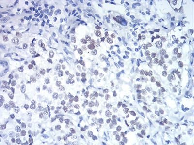
Figure 6:Immunohistochemical analysis of paraffin-embedded bladder cancer tissues using DDX5 mouse mAb with DAB staining.
For Research Use Only. Not for use in diagnostic procedures.