

DDX39B Primary Antibody
Item Information
Catalog #
Size
Price
Description
This gene encodes a member of the DEAD box family of RNA-dependent ATPases that mediate ATP hydrolysis during pre-mRNA splicing. The encoded protein is an essential splicing factor required for association of U2 small nuclear ribonucleoprotein with pre-mRNA, and it also plays an important role in mRNA export from the nucleus to the cytoplasm. This gene belongs to a cluster of genes localized in the vicinity of the genes encoding tumor necrosis factor alpha and tumor necrosis factor beta. These genes are all within the human major histocompatibility complex class III region. Mutations in this gene may be associated with rheumatoid arthritis. Alternative splicing results in multiple transcript variants. Related pseudogenes have been identified on both chromosomes 6 and 11. Read-through transcription also occurs between this gene and the upstream ATP6V1G2 (ATPase, H+ transporting, lysosomal 13kDa, V1 subunit G2) gene.
Product Overview
Entrez GenelD
7919
Aliases
BAT1; UAP56; D6S81E
Clone#
3A2B2
Host / Isotype
Mouse / IgG1
Species Reactivity
Human, Mouse
Immunogen
Purified recombinant fragment of human DDX39B (AA: 1-250) expressed in E. Coli.
Formulation
Purified antibody in PBS with 0.05% sodium azide
Storage
Store at 4°C short term. Aliquot and store at -20°C long term. Avoid freeze/thaw cycles.
Product Applications
WB (Western Blot)
1/500 - 1/2000
IHC_P(Immunohistochemistry)
1/200 - 1/1000
ELISA
1/10000
References
1.J Virol. 2011 Sep;85(17):8646-55.
2.Biochem Biophys Res Commun. 2010 Feb 26;393(1):106-10.
2.Biochem Biophys Res Commun. 2010 Feb 26;393(1):106-10.
Product Image
Elisa
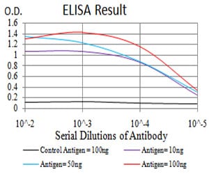
Figure 1: Black line: Control Antigen (100 ng); Purple line: Antigen(10ng); Blue line: Antigen (50 ng); Red line: Antigen (100 ng);
Western Blot
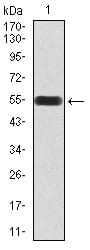
Figure 2:Western blot analysis using DDX39B mAb against human DDX39B (AA: 1-250) recombinant protein. (Expected MW is 54.2 kDa)
Western Blot

Figure 3:Western blot analysis using DDX39B mAb against HEK293 (1) and DDX39B (AA: 1-250)-hIgGFc transfected HEK293 (2) cell lysate.
Immunohistochemical analysis

Figure 5:Immunohistochemical analysis of paraffin-embedded cervical cancer tissues using DDX39B mouse mAb with DAB staining.
Western Blot

Figure 6:Western blot analysis using DDX39B mouse mAb against HEK293 (1), Jurkat (2), MCF-7 (3), A431 (4), NIH/3T3 (5), Jurkat (6), K562 (7), and HepG2 (8) cell lysate.
For Research Use Only. Not for use in diagnostic procedures.